Magnesium »
PDB 3ism-3jaw »
3itu »
Magnesium in PDB 3itu: HPDE2A Catalytic Domain Complexed with Ibmx
Enzymatic activity of HPDE2A Catalytic Domain Complexed with Ibmx
All present enzymatic activity of HPDE2A Catalytic Domain Complexed with Ibmx:
3.1.4.17;
3.1.4.17;
Protein crystallography data
The structure of HPDE2A Catalytic Domain Complexed with Ibmx, PDB code: 3itu
was solved by
J.Pandit,
with X-Ray Crystallography technique. A brief refinement statistics is given in the table below:
| Resolution Low / High (Å) | 50.00 / 1.58 |
| Space group | P 1 |
| Cell size a, b, c (Å), α, β, γ (°) | 55.815, 73.287, 91.532, 109.30, 88.80, 89.00 |
| R / Rfree (%) | 17.4 / 23.3 |
Other elements in 3itu:
The structure of HPDE2A Catalytic Domain Complexed with Ibmx also contains other interesting chemical elements:
| Zinc | (Zn) | 4 atoms |
Magnesium Binding Sites:
The binding sites of Magnesium atom in the HPDE2A Catalytic Domain Complexed with Ibmx
(pdb code 3itu). This binding sites where shown within
5.0 Angstroms radius around Magnesium atom.
In total 4 binding sites of Magnesium where determined in the HPDE2A Catalytic Domain Complexed with Ibmx, PDB code: 3itu:
Jump to Magnesium binding site number: 1; 2; 3; 4;
In total 4 binding sites of Magnesium where determined in the HPDE2A Catalytic Domain Complexed with Ibmx, PDB code: 3itu:
Jump to Magnesium binding site number: 1; 2; 3; 4;
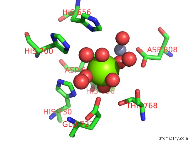
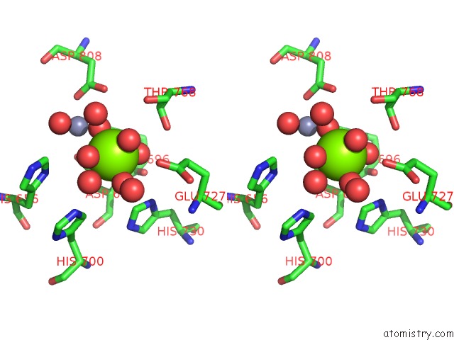
Magnesium binding site 1 out of 4 in 3itu
Go back to
Magnesium binding site 1 out
of 4 in the HPDE2A Catalytic Domain Complexed with Ibmx
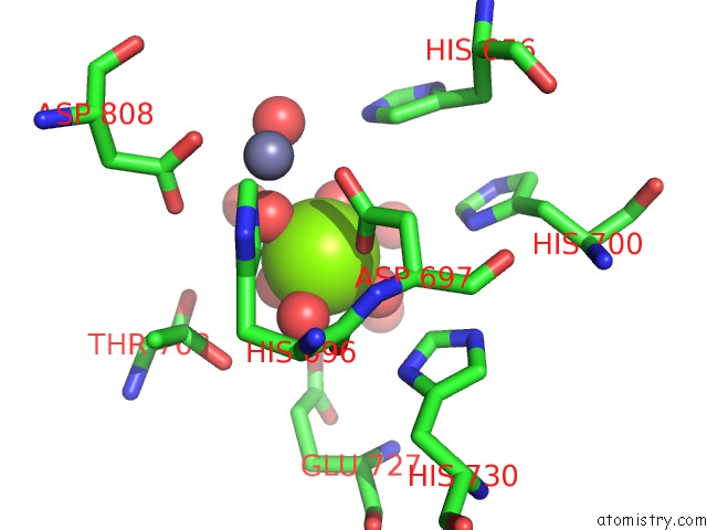
Mono view
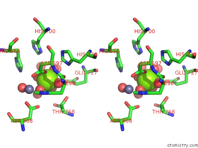
Stereo pair view
Mono view
Stereo pair view
A full contact list of Magnesium with other atoms in the Mg binding
site number 1 of HPDE2A Catalytic Domain Complexed with Ibmx within 5.0Å range:
|
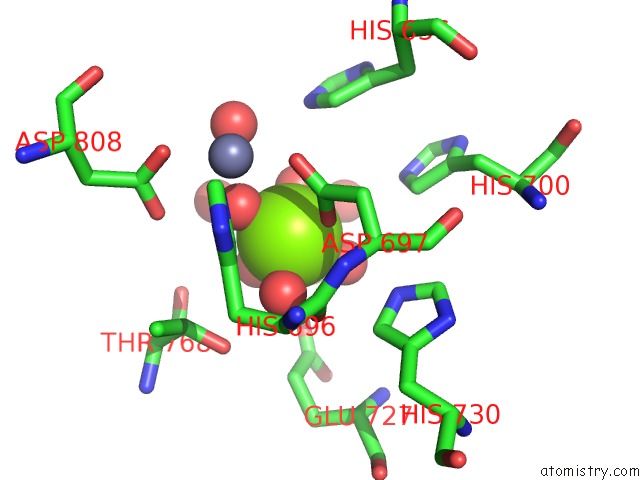
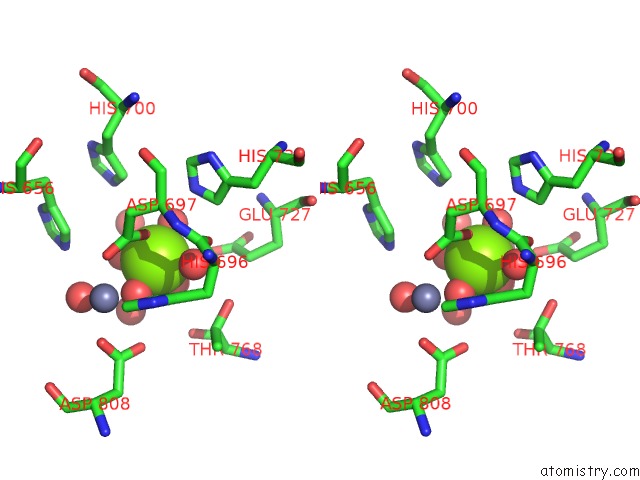
Magnesium binding site 2 out of 4 in 3itu
Go back to
Magnesium binding site 2 out
of 4 in the HPDE2A Catalytic Domain Complexed with Ibmx
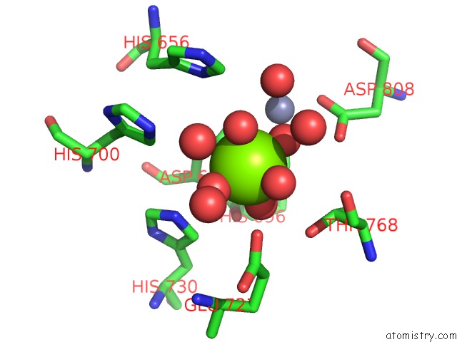
Mono view
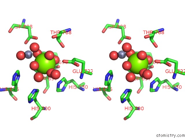
Stereo pair view
Mono view
Stereo pair view
A full contact list of Magnesium with other atoms in the Mg binding
site number 2 of HPDE2A Catalytic Domain Complexed with Ibmx within 5.0Å range:
|
Magnesium binding site 3 out of 4 in 3itu
Go back to
Magnesium binding site 3 out
of 4 in the HPDE2A Catalytic Domain Complexed with Ibmx
Mono view
Stereo pair view
Mono view
Stereo pair view
A full contact list of Magnesium with other atoms in the Mg binding
site number 3 of HPDE2A Catalytic Domain Complexed with Ibmx within 5.0Å range:
|
Magnesium binding site 4 out of 4 in 3itu
Go back to
Magnesium binding site 4 out
of 4 in the HPDE2A Catalytic Domain Complexed with Ibmx
Mono view
Stereo pair view
Mono view
Stereo pair view
A full contact list of Magnesium with other atoms in the Mg binding
site number 4 of HPDE2A Catalytic Domain Complexed with Ibmx within 5.0Å range:
|
Reference:
J.Pandit,
M.D.Forman,
K.F.Fennell,
K.S.Dillman,
F.S.Menniti.
Mechanism For the Allosteric Regulation of Phosphodiesterase 2A Deduced From the X-Ray Structure of A Near Full-Length Construct. Proc.Natl.Acad.Sci.Usa V. 106 18225 2009.
ISSN: ISSN 0027-8424
PubMed: 19828435
DOI: 10.1073/PNAS.0907635106
Page generated: Sun Aug 10 22:42:45 2025
ISSN: ISSN 0027-8424
PubMed: 19828435
DOI: 10.1073/PNAS.0907635106
Last articles
Na in 9MM2Na in 9LR7
Na in 9MBL
Na in 9ME8
Na in 9MBK
Na in 9MBH
Na in 9MBG
Na in 9MBE
Na in 9MBF
Na in 9M56