Magnesium »
PDB 3ism-3jaw »
3ivk »
Magnesium in PDB 3ivk: Crystal Structure of the Catalytic Core of An Rna Polymerase Ribozyme Complexed with An Antigen Binding Antibody Fragment
Protein crystallography data
The structure of Crystal Structure of the Catalytic Core of An Rna Polymerase Ribozyme Complexed with An Antigen Binding Antibody Fragment, PDB code: 3ivk
was solved by
Y.Koldobskaya,
E.M.Duguid,
D.M.Shechner,
S.Koide,
A.A.Kossiakoff,
D.P.Bartel,
J.A.Piccirilli,
with X-Ray Crystallography technique. A brief refinement statistics is given in the table below:
| Resolution Low / High (Å) | 50.00 / 3.10 |
| Space group | C 2 2 21 |
| Cell size a, b, c (Å), α, β, γ (°) | 207.499, 206.452, 135.926, 90.00, 90.00, 90.00 |
| R / Rfree (%) | 20.7 / 22.5 |
Other elements in 3ivk:
The structure of Crystal Structure of the Catalytic Core of An Rna Polymerase Ribozyme Complexed with An Antigen Binding Antibody Fragment also contains other interesting chemical elements:
| Cadmium | (Cd) | 2 atoms |
| Chlorine | (Cl) | 2 atoms |
Magnesium Binding Sites:
Pages:
>>> Page 1 <<< Page 2, Binding sites: 11 - 20; Page 3, Binding sites: 21 - 30; Page 4, Binding sites: 31 - 33;Binding sites:
The binding sites of Magnesium atom in the Crystal Structure of the Catalytic Core of An Rna Polymerase Ribozyme Complexed with An Antigen Binding Antibody Fragment (pdb code 3ivk). This binding sites where shown within 5.0 Angstroms radius around Magnesium atom.In total 33 binding sites of Magnesium where determined in the Crystal Structure of the Catalytic Core of An Rna Polymerase Ribozyme Complexed with An Antigen Binding Antibody Fragment, PDB code: 3ivk:
Jump to Magnesium binding site number: 1; 2; 3; 4; 5; 6; 7; 8; 9; 10;
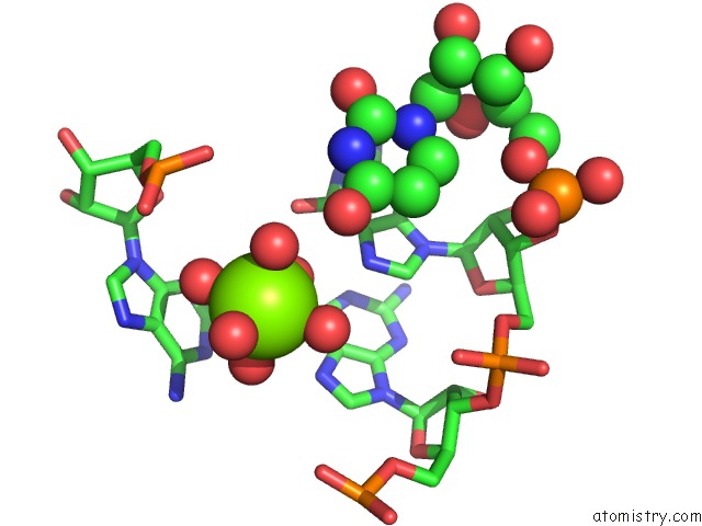
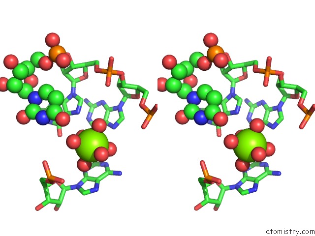
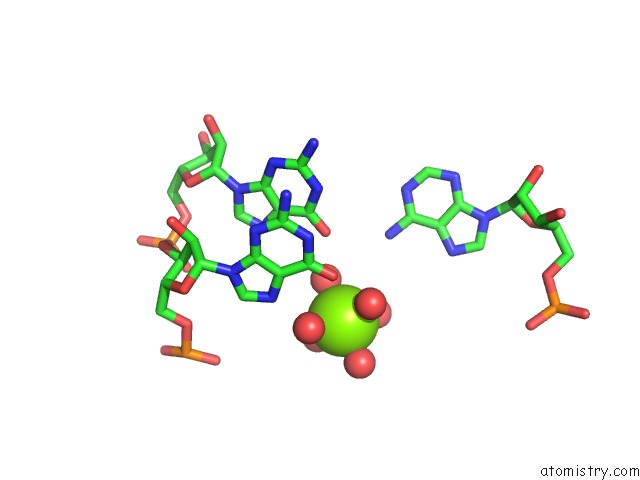
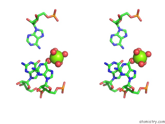
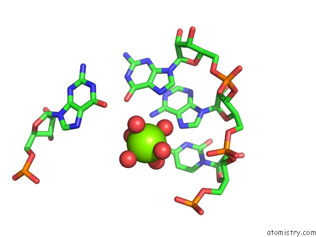
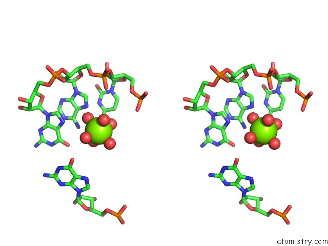
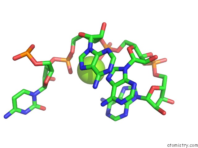
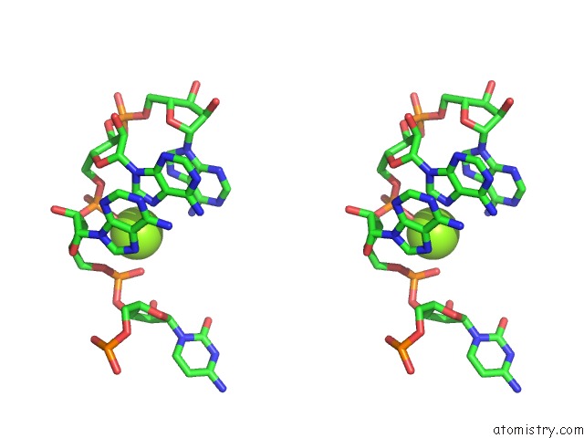
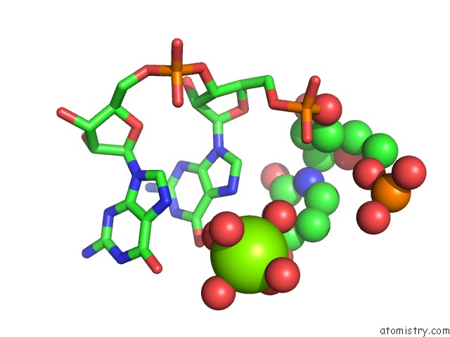
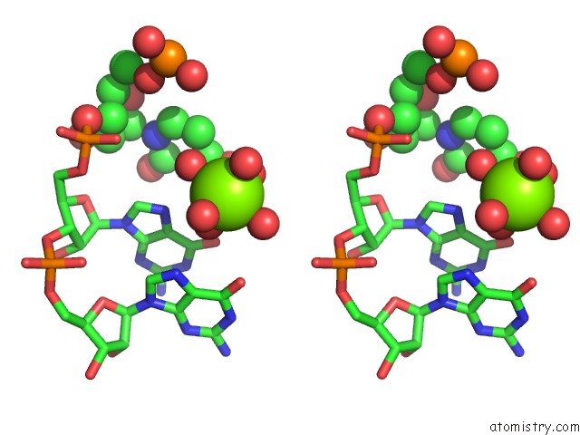
Magnesium binding site 1 out of 33 in 3ivk
Go back to
Magnesium binding site 1 out
of 33 in the Crystal Structure of the Catalytic Core of An Rna Polymerase Ribozyme Complexed with An Antigen Binding Antibody Fragment
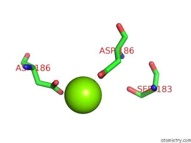
Mono view
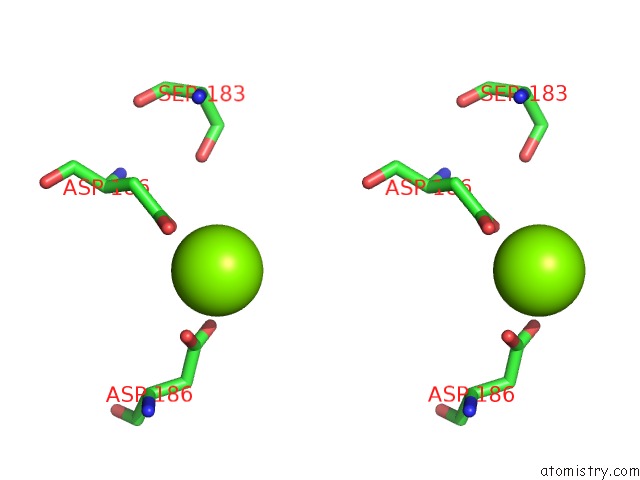
Stereo pair view
Mono view
Stereo pair view
A full contact list of Magnesium with other atoms in the Mg binding
site number 1 of Crystal Structure of the Catalytic Core of An Rna Polymerase Ribozyme Complexed with An Antigen Binding Antibody Fragment within 5.0Å range:
|
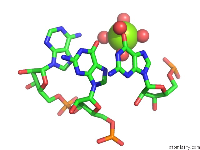
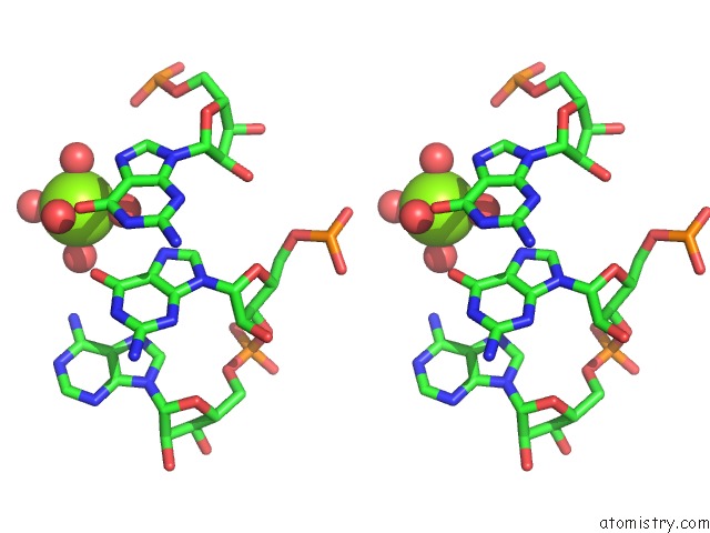
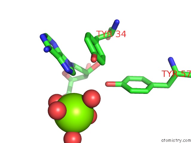
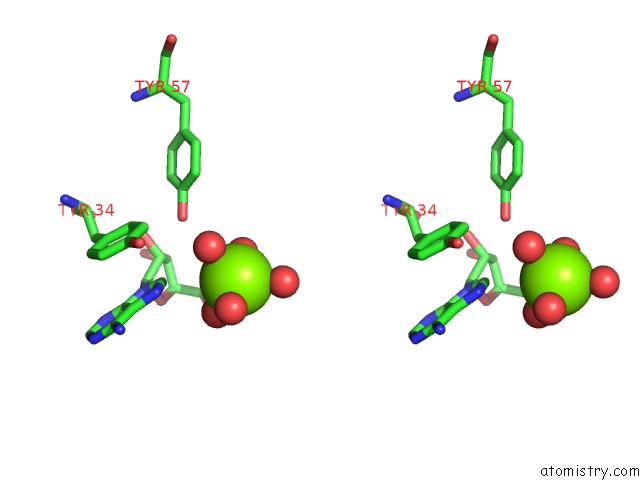
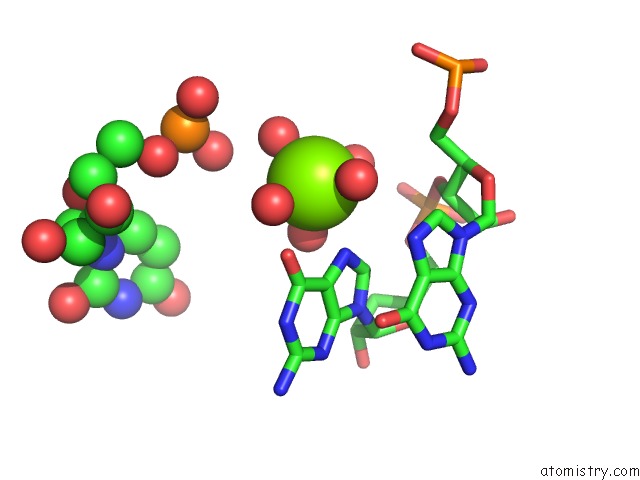
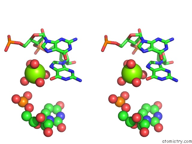
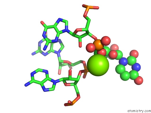
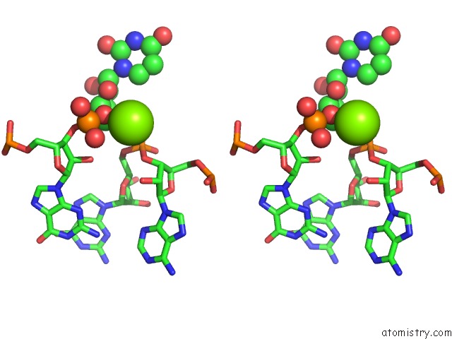
Magnesium binding site 2 out of 33 in 3ivk
Go back to
Magnesium binding site 2 out
of 33 in the Crystal Structure of the Catalytic Core of An Rna Polymerase Ribozyme Complexed with An Antigen Binding Antibody Fragment
Mono view
Stereo pair view
Mono view
Stereo pair view
A full contact list of Magnesium with other atoms in the Mg binding
site number 2 of Crystal Structure of the Catalytic Core of An Rna Polymerase Ribozyme Complexed with An Antigen Binding Antibody Fragment within 5.0Å range:
|
Magnesium binding site 3 out of 33 in 3ivk
Go back to
Magnesium binding site 3 out
of 33 in the Crystal Structure of the Catalytic Core of An Rna Polymerase Ribozyme Complexed with An Antigen Binding Antibody Fragment
Mono view
Stereo pair view
Mono view
Stereo pair view
A full contact list of Magnesium with other atoms in the Mg binding
site number 3 of Crystal Structure of the Catalytic Core of An Rna Polymerase Ribozyme Complexed with An Antigen Binding Antibody Fragment within 5.0Å range:
|
Magnesium binding site 4 out of 33 in 3ivk
Go back to
Magnesium binding site 4 out
of 33 in the Crystal Structure of the Catalytic Core of An Rna Polymerase Ribozyme Complexed with An Antigen Binding Antibody Fragment
Mono view
Stereo pair view
Mono view
Stereo pair view
A full contact list of Magnesium with other atoms in the Mg binding
site number 4 of Crystal Structure of the Catalytic Core of An Rna Polymerase Ribozyme Complexed with An Antigen Binding Antibody Fragment within 5.0Å range:
|
Magnesium binding site 5 out of 33 in 3ivk
Go back to
Magnesium binding site 5 out
of 33 in the Crystal Structure of the Catalytic Core of An Rna Polymerase Ribozyme Complexed with An Antigen Binding Antibody Fragment
Mono view
Stereo pair view
Mono view
Stereo pair view
A full contact list of Magnesium with other atoms in the Mg binding
site number 5 of Crystal Structure of the Catalytic Core of An Rna Polymerase Ribozyme Complexed with An Antigen Binding Antibody Fragment within 5.0Å range:
|
Magnesium binding site 6 out of 33 in 3ivk
Go back to
Magnesium binding site 6 out
of 33 in the Crystal Structure of the Catalytic Core of An Rna Polymerase Ribozyme Complexed with An Antigen Binding Antibody Fragment
Mono view
Stereo pair view
Mono view
Stereo pair view
A full contact list of Magnesium with other atoms in the Mg binding
site number 6 of Crystal Structure of the Catalytic Core of An Rna Polymerase Ribozyme Complexed with An Antigen Binding Antibody Fragment within 5.0Å range:
|
Magnesium binding site 7 out of 33 in 3ivk
Go back to
Magnesium binding site 7 out
of 33 in the Crystal Structure of the Catalytic Core of An Rna Polymerase Ribozyme Complexed with An Antigen Binding Antibody Fragment
Mono view
Stereo pair view
Mono view
Stereo pair view
A full contact list of Magnesium with other atoms in the Mg binding
site number 7 of Crystal Structure of the Catalytic Core of An Rna Polymerase Ribozyme Complexed with An Antigen Binding Antibody Fragment within 5.0Å range:
|
Magnesium binding site 8 out of 33 in 3ivk
Go back to
Magnesium binding site 8 out
of 33 in the Crystal Structure of the Catalytic Core of An Rna Polymerase Ribozyme Complexed with An Antigen Binding Antibody Fragment
Mono view
Stereo pair view
Mono view
Stereo pair view
A full contact list of Magnesium with other atoms in the Mg binding
site number 8 of Crystal Structure of the Catalytic Core of An Rna Polymerase Ribozyme Complexed with An Antigen Binding Antibody Fragment within 5.0Å range:
|
Magnesium binding site 9 out of 33 in 3ivk
Go back to
Magnesium binding site 9 out
of 33 in the Crystal Structure of the Catalytic Core of An Rna Polymerase Ribozyme Complexed with An Antigen Binding Antibody Fragment
Mono view
Stereo pair view
Mono view
Stereo pair view
A full contact list of Magnesium with other atoms in the Mg binding
site number 9 of Crystal Structure of the Catalytic Core of An Rna Polymerase Ribozyme Complexed with An Antigen Binding Antibody Fragment within 5.0Å range:
|
Magnesium binding site 10 out of 33 in 3ivk
Go back to
Magnesium binding site 10 out
of 33 in the Crystal Structure of the Catalytic Core of An Rna Polymerase Ribozyme Complexed with An Antigen Binding Antibody Fragment
Mono view
Stereo pair view
Mono view
Stereo pair view
A full contact list of Magnesium with other atoms in the Mg binding
site number 10 of Crystal Structure of the Catalytic Core of An Rna Polymerase Ribozyme Complexed with An Antigen Binding Antibody Fragment within 5.0Å range:
|
Reference:
D.M.Shechner,
R.A.Grant,
S.C.Bagby,
Y.Koldobskaya,
J.A.Piccirilli,
D.P.Bartel.
Crystal Structure of the Catalytic Core of An Rna-Polymerase Ribozyme. Science V. 326 1271 2009.
ISSN: ISSN 0036-8075
PubMed: 19965478
DOI: 10.1126/SCIENCE.1174676
Page generated: Wed Aug 14 16:19:41 2024
ISSN: ISSN 0036-8075
PubMed: 19965478
DOI: 10.1126/SCIENCE.1174676
Last articles
K in 1D7UK in 1D7S
K in 1D7R
K in 1C38
K in 1CPG
K in 1CPE
K in 1C30
K in 1C7J
K in 1C35
K in 1BXR