Magnesium »
PDB 3jay-3jwr »
3ju8 »
Magnesium in PDB 3ju8: Crystal Structure of Succinylglutamic Semialdehyde Dehydrogenase From Pseudomonas Aeruginosa.
Enzymatic activity of Crystal Structure of Succinylglutamic Semialdehyde Dehydrogenase From Pseudomonas Aeruginosa.
All present enzymatic activity of Crystal Structure of Succinylglutamic Semialdehyde Dehydrogenase From Pseudomonas Aeruginosa.:
1.2.1.71;
1.2.1.71;
Protein crystallography data
The structure of Crystal Structure of Succinylglutamic Semialdehyde Dehydrogenase From Pseudomonas Aeruginosa., PDB code: 3ju8
was solved by
Y.Kim,
H.Li,
K.Buck,
A.Joachimiak,
Midwest Center For Structural Genomics(Mcsg),
with X-Ray Crystallography technique. A brief refinement statistics is given in the table below:
| Resolution Low / High (Å) | 32.77 / 1.82 |
| Space group | C 1 2 1 |
| Cell size a, b, c (Å), α, β, γ (°) | 137.117, 48.033, 187.130, 90.00, 107.43, 90.00 |
| R / Rfree (%) | 15.8 / 19.6 |
Other elements in 3ju8:
The structure of Crystal Structure of Succinylglutamic Semialdehyde Dehydrogenase From Pseudomonas Aeruginosa. also contains other interesting chemical elements:
| Chlorine | (Cl) | 2 atoms |
Magnesium Binding Sites:
The binding sites of Magnesium atom in the Crystal Structure of Succinylglutamic Semialdehyde Dehydrogenase From Pseudomonas Aeruginosa.
(pdb code 3ju8). This binding sites where shown within
5.0 Angstroms radius around Magnesium atom.
In total only one binding site of Magnesium was determined in the Crystal Structure of Succinylglutamic Semialdehyde Dehydrogenase From Pseudomonas Aeruginosa., PDB code: 3ju8:
In total only one binding site of Magnesium was determined in the Crystal Structure of Succinylglutamic Semialdehyde Dehydrogenase From Pseudomonas Aeruginosa., PDB code: 3ju8:
Magnesium binding site 1 out of 1 in 3ju8
Go back to
Magnesium binding site 1 out
of 1 in the Crystal Structure of Succinylglutamic Semialdehyde Dehydrogenase From Pseudomonas Aeruginosa.
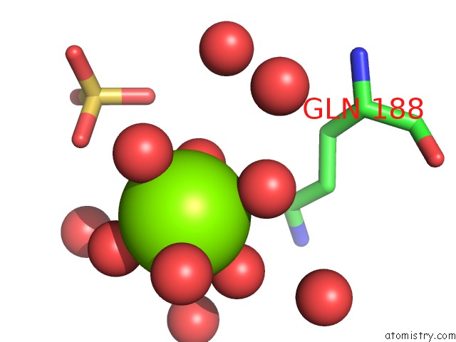
Mono view
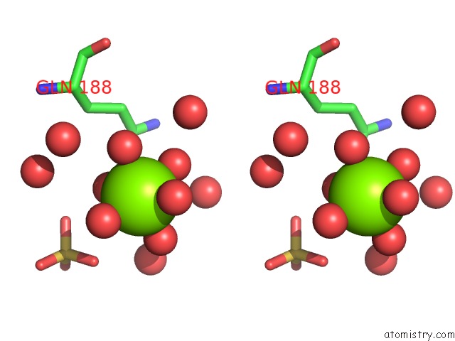
Stereo pair view
Mono view
Stereo pair view
A full contact list of Magnesium with other atoms in the Mg binding
site number 1 of Crystal Structure of Succinylglutamic Semialdehyde Dehydrogenase From Pseudomonas Aeruginosa. within 5.0Å range:
|
Reference:
Y.Kim,
H.Li,
K.Buck,
A.Joachimiak.
Crystal Structure of Succinylglutamic Semialdehyde Dehydrogenase From Pseudomonas Aeruginosa To Be Published.
Page generated: Wed Aug 14 17:13:45 2024
Last articles
Mg in 1GXBMg in 1H1D
Mg in 1GZG
Mg in 1H17
Mg in 1GY3
Mg in 1H16
Mg in 1GUT
Mg in 1GWN
Mg in 1GTV
Mg in 1GUS