Magnesium »
PDB 3ktv-3l8z »
3kws »
Magnesium in PDB 3kws: Crystal Structure of Putative Sugar Isomerase (YP_001305149.1) From Parabacteroides Distasonis Atcc 8503 at 1.68 A Resolution
Protein crystallography data
The structure of Crystal Structure of Putative Sugar Isomerase (YP_001305149.1) From Parabacteroides Distasonis Atcc 8503 at 1.68 A Resolution, PDB code: 3kws
was solved by
Joint Center For Structural Genomics (Jcsg),
with X-Ray Crystallography technique. A brief refinement statistics is given in the table below:
| Resolution Low / High (Å) | 29.37 / 1.68 |
| Space group | P 21 21 21 |
| Cell size a, b, c (Å), α, β, γ (°) | 51.573, 107.200, 113.146, 90.00, 90.00, 90.00 |
| R / Rfree (%) | 15 / 18.2 |
Magnesium Binding Sites:
The binding sites of Magnesium atom in the Crystal Structure of Putative Sugar Isomerase (YP_001305149.1) From Parabacteroides Distasonis Atcc 8503 at 1.68 A Resolution
(pdb code 3kws). This binding sites where shown within
5.0 Angstroms radius around Magnesium atom.
In total 2 binding sites of Magnesium where determined in the Crystal Structure of Putative Sugar Isomerase (YP_001305149.1) From Parabacteroides Distasonis Atcc 8503 at 1.68 A Resolution, PDB code: 3kws:
Jump to Magnesium binding site number: 1; 2;
In total 2 binding sites of Magnesium where determined in the Crystal Structure of Putative Sugar Isomerase (YP_001305149.1) From Parabacteroides Distasonis Atcc 8503 at 1.68 A Resolution, PDB code: 3kws:
Jump to Magnesium binding site number: 1; 2;
Magnesium binding site 1 out of 2 in 3kws
Go back to
Magnesium binding site 1 out
of 2 in the Crystal Structure of Putative Sugar Isomerase (YP_001305149.1) From Parabacteroides Distasonis Atcc 8503 at 1.68 A Resolution
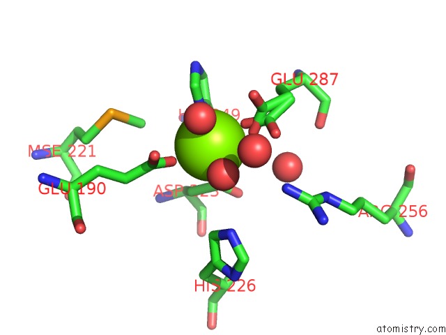
Mono view
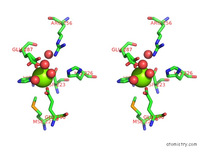
Stereo pair view
Mono view
Stereo pair view
A full contact list of Magnesium with other atoms in the Mg binding
site number 1 of Crystal Structure of Putative Sugar Isomerase (YP_001305149.1) From Parabacteroides Distasonis Atcc 8503 at 1.68 A Resolution within 5.0Å range:
|
Magnesium binding site 2 out of 2 in 3kws
Go back to
Magnesium binding site 2 out
of 2 in the Crystal Structure of Putative Sugar Isomerase (YP_001305149.1) From Parabacteroides Distasonis Atcc 8503 at 1.68 A Resolution
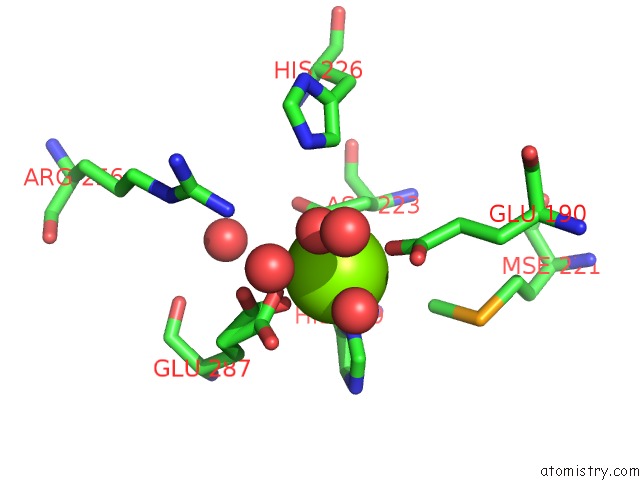
Mono view
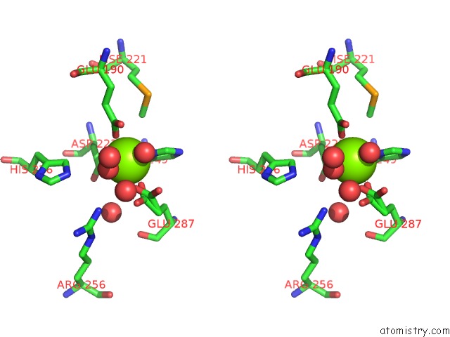
Stereo pair view
Mono view
Stereo pair view
A full contact list of Magnesium with other atoms in the Mg binding
site number 2 of Crystal Structure of Putative Sugar Isomerase (YP_001305149.1) From Parabacteroides Distasonis Atcc 8503 at 1.68 A Resolution within 5.0Å range:
|
Reference:
Joint Center For Structural Genomics (Jcsg),
Joint Center For Structural Genomics (Jcsg).
N/A N/A.
Page generated: Wed Aug 14 18:18:14 2024
Last articles
Ca in 6LERCa in 6LGB
Ca in 6LA9
Ca in 6LGA
Ca in 6LFJ
Ca in 6LF7
Ca in 6LEH
Ca in 6LDY
Ca in 6LB7
Ca in 6LAB