Magnesium »
PDB 3m1v-3mfh »
3mbo »
Magnesium in PDB 3mbo: Crystal Structure of the Glycosyltransferase Babsha Bound with Udp and L-Malate
Protein crystallography data
The structure of Crystal Structure of the Glycosyltransferase Babsha Bound with Udp and L-Malate, PDB code: 3mbo
was solved by
B.D.Wallace,
A.Claiborne,
M.R.Redinbo,
with X-Ray Crystallography technique. A brief refinement statistics is given in the table below:
| Resolution Low / High (Å) | 48.22 / 3.31 |
| Space group | P 41 |
| Cell size a, b, c (Å), α, β, γ (°) | 226.266, 226.266, 75.354, 90.00, 90.00, 90.00 |
| R / Rfree (%) | 22.8 / 25.7 |
Magnesium Binding Sites:
The binding sites of Magnesium atom in the Crystal Structure of the Glycosyltransferase Babsha Bound with Udp and L-Malate
(pdb code 3mbo). This binding sites where shown within
5.0 Angstroms radius around Magnesium atom.
In total 2 binding sites of Magnesium where determined in the Crystal Structure of the Glycosyltransferase Babsha Bound with Udp and L-Malate, PDB code: 3mbo:
Jump to Magnesium binding site number: 1; 2;
In total 2 binding sites of Magnesium where determined in the Crystal Structure of the Glycosyltransferase Babsha Bound with Udp and L-Malate, PDB code: 3mbo:
Jump to Magnesium binding site number: 1; 2;
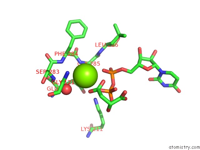
Magnesium binding site 1 out of 2 in 3mbo
Go back to
Magnesium binding site 1 out
of 2 in the Crystal Structure of the Glycosyltransferase Babsha Bound with Udp and L-Malate
Mono view

Stereo pair view
Mono view
Stereo pair view
A full contact list of Magnesium with other atoms in the Mg binding
site number 1 of Crystal Structure of the Glycosyltransferase Babsha Bound with Udp and L-Malate within 5.0Å range:
|
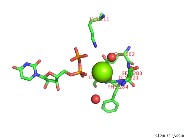
Magnesium binding site 2 out of 2 in 3mbo
Go back to
Magnesium binding site 2 out
of 2 in the Crystal Structure of the Glycosyltransferase Babsha Bound with Udp and L-Malate
Mono view

Stereo pair view
Mono view
Stereo pair view
A full contact list of Magnesium with other atoms in the Mg binding
site number 2 of Crystal Structure of the Glycosyltransferase Babsha Bound with Udp and L-Malate within 5.0Å range:
|
Reference:
D.Parsonage,
G.L.Newton,
R.C.Holder,
B.D.Wallace,
C.Paige,
P.C.Dos Santos,
M.R.Redinbo,
R.C.Fahey,
S.D.Reid,
A.Claiborne.
Characterization of the N-Acetyl-Alpha-D-Glucosaminyl L-Malate Synthase and Deacetylase Functions For Bacillithiol Biosynthesis in Bacillus Anthracis Biochemistry V. 49 8398 2010.
ISSN: ISSN 0006-2960
Page generated: Mon Aug 11 00:31:13 2025
ISSN: ISSN 0006-2960
Last articles
Mg in 5GQJMg in 5GL3
Mg in 5GQI
Mg in 5GMK
Mg in 5GON
Mg in 5GPA
Mg in 5GP9
Mg in 5GOF
Mg in 5GMY
Mg in 5GOD