Magnesium »
PDB 3mv1-3n8b »
3my9 »
Magnesium in PDB 3my9: Crystal Structure of A Muconate Cycloisomerase From Azorhizobium Caulinodans
Protein crystallography data
The structure of Crystal Structure of A Muconate Cycloisomerase From Azorhizobium Caulinodans, PDB code: 3my9
was solved by
C.E.Quartararo,
U.Ramagopal,
J.B.Bonanno,
M.Rutter,
K.T.Bain,
S.Miller,
R.Toro,
J.M.Sauder,
S.K.Burley,
S.C.Almo,
New York Sgx Research Centerfor Structural Genomics (Nysgxrc),
with X-Ray Crystallography technique. A brief refinement statistics is given in the table below:
| Resolution Low / High (Å) | 20.00 / 2.20 |
| Space group | I 4 2 2 |
| Cell size a, b, c (Å), α, β, γ (°) | 127.993, 127.993, 98.036, 90.00, 90.00, 90.00 |
| R / Rfree (%) | 20.2 / 25.4 |
Magnesium Binding Sites:
The binding sites of Magnesium atom in the Crystal Structure of A Muconate Cycloisomerase From Azorhizobium Caulinodans
(pdb code 3my9). This binding sites where shown within
5.0 Angstroms radius around Magnesium atom.
In total 2 binding sites of Magnesium where determined in the Crystal Structure of A Muconate Cycloisomerase From Azorhizobium Caulinodans, PDB code: 3my9:
Jump to Magnesium binding site number: 1; 2;
In total 2 binding sites of Magnesium where determined in the Crystal Structure of A Muconate Cycloisomerase From Azorhizobium Caulinodans, PDB code: 3my9:
Jump to Magnesium binding site number: 1; 2;
Magnesium binding site 1 out of 2 in 3my9
Go back to
Magnesium binding site 1 out
of 2 in the Crystal Structure of A Muconate Cycloisomerase From Azorhizobium Caulinodans
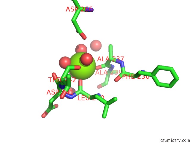
Mono view
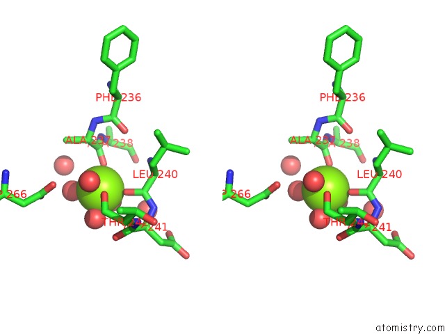
Stereo pair view
Mono view
Stereo pair view
A full contact list of Magnesium with other atoms in the Mg binding
site number 1 of Crystal Structure of A Muconate Cycloisomerase From Azorhizobium Caulinodans within 5.0Å range:
|
Magnesium binding site 2 out of 2 in 3my9
Go back to
Magnesium binding site 2 out
of 2 in the Crystal Structure of A Muconate Cycloisomerase From Azorhizobium Caulinodans
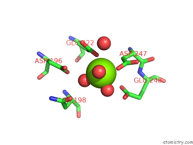
Mono view
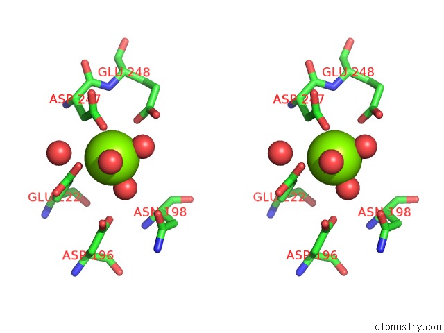
Stereo pair view
Mono view
Stereo pair view
A full contact list of Magnesium with other atoms in the Mg binding
site number 2 of Crystal Structure of A Muconate Cycloisomerase From Azorhizobium Caulinodans within 5.0Å range:
|
Reference:
C.E.Quartararo,
U.Ramagopal,
J.B.Bonanno,
M.Rutter,
K.T.Bain,
S.Miller,
R.Toro,
J.M.Sauder,
S.K.Burley,
S.C.Almo.
Crystal Structure of A Muconate Cycloisomerase From Azorhizobium Caulinodans To Be Published.
Page generated: Mon Aug 11 00:44:00 2025
Last articles
Mg in 5T14Mg in 5SZT
Mg in 5T2V
Mg in 5T13
Mg in 5T19
Mg in 5SZK
Mg in 5SZJ
Mg in 5SZI
Mg in 5SZH
Mg in 5SWD