Magnesium »
PDB 3nl0-3o0q »
3no1 »
Magnesium in PDB 3no1: Crystal Structure of Mandelate Racemase/Muconate Lactonizing Enzyme From A Marine Actinobacterium in Complex with Magnesium
Protein crystallography data
The structure of Crystal Structure of Mandelate Racemase/Muconate Lactonizing Enzyme From A Marine Actinobacterium in Complex with Magnesium, PDB code: 3no1
was solved by
L.Satyanarayana,
S.K.Burley,
S.Swaminathan,
New York Sgx Research Centerfor Structural Genomics (Nysgxrc),
with X-Ray Crystallography technique. A brief refinement statistics is given in the table below:
| Resolution Low / High (Å) | 42.51 / 2.16 |
| Space group | P 1 |
| Cell size a, b, c (Å), α, β, γ (°) | 84.961, 90.103, 94.660, 115.80, 109.75, 97.58 |
| R / Rfree (%) | 20.7 / 24 |
Magnesium Binding Sites:
The binding sites of Magnesium atom in the Crystal Structure of Mandelate Racemase/Muconate Lactonizing Enzyme From A Marine Actinobacterium in Complex with Magnesium
(pdb code 3no1). This binding sites where shown within
5.0 Angstroms radius around Magnesium atom.
In total 6 binding sites of Magnesium where determined in the Crystal Structure of Mandelate Racemase/Muconate Lactonizing Enzyme From A Marine Actinobacterium in Complex with Magnesium, PDB code: 3no1:
Jump to Magnesium binding site number: 1; 2; 3; 4; 5; 6;
In total 6 binding sites of Magnesium where determined in the Crystal Structure of Mandelate Racemase/Muconate Lactonizing Enzyme From A Marine Actinobacterium in Complex with Magnesium, PDB code: 3no1:
Jump to Magnesium binding site number: 1; 2; 3; 4; 5; 6;
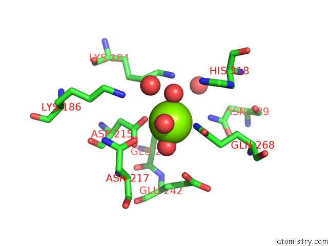
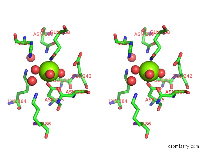
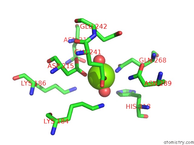
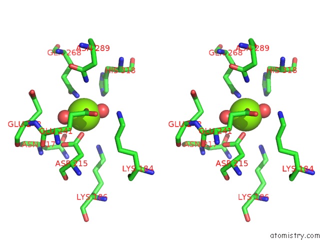
Magnesium binding site 1 out of 6 in 3no1
Go back to
Magnesium binding site 1 out
of 6 in the Crystal Structure of Mandelate Racemase/Muconate Lactonizing Enzyme From A Marine Actinobacterium in Complex with Magnesium
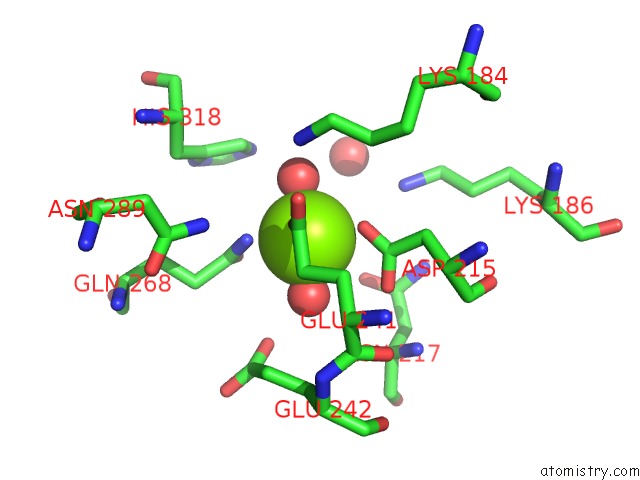
Mono view
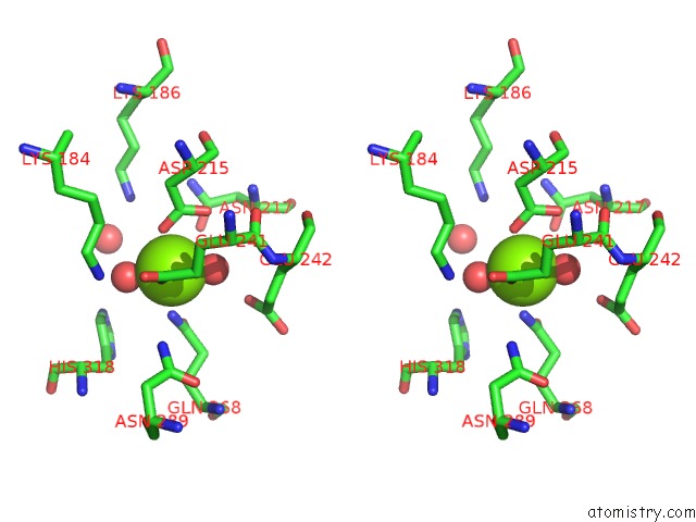
Stereo pair view
Mono view
Stereo pair view
A full contact list of Magnesium with other atoms in the Mg binding
site number 1 of Crystal Structure of Mandelate Racemase/Muconate Lactonizing Enzyme From A Marine Actinobacterium in Complex with Magnesium within 5.0Å range:
|
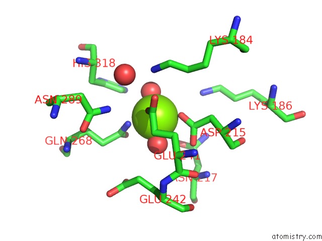
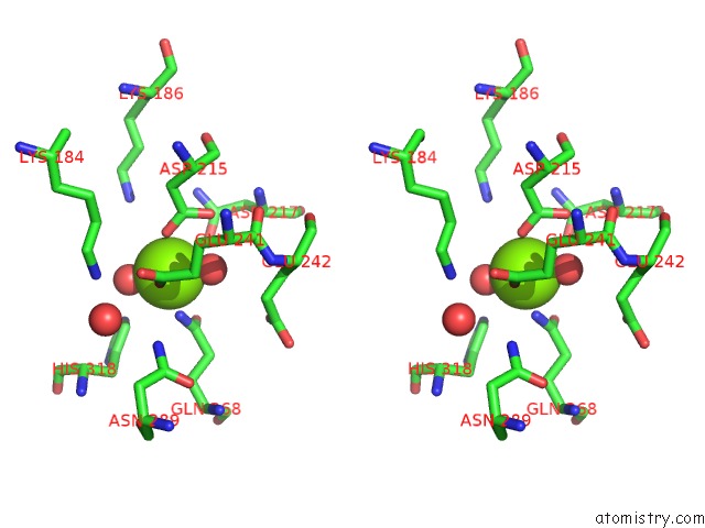
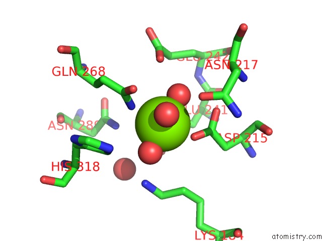
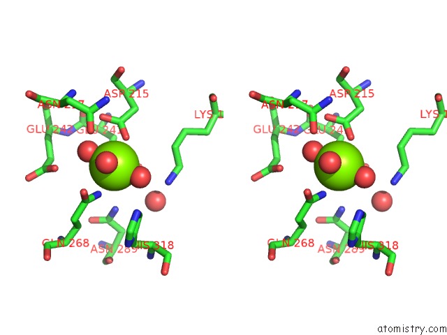
Magnesium binding site 2 out of 6 in 3no1
Go back to
Magnesium binding site 2 out
of 6 in the Crystal Structure of Mandelate Racemase/Muconate Lactonizing Enzyme From A Marine Actinobacterium in Complex with Magnesium
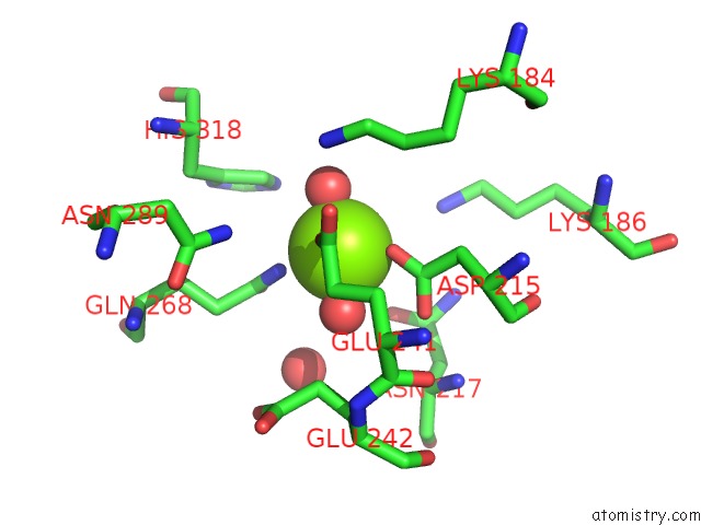
Mono view
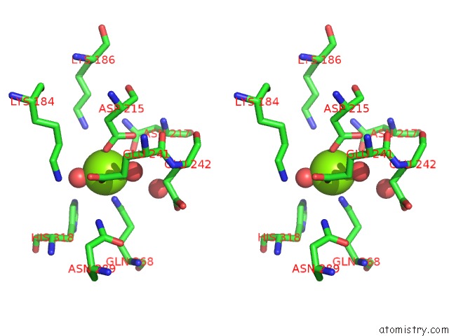
Stereo pair view
Mono view
Stereo pair view
A full contact list of Magnesium with other atoms in the Mg binding
site number 2 of Crystal Structure of Mandelate Racemase/Muconate Lactonizing Enzyme From A Marine Actinobacterium in Complex with Magnesium within 5.0Å range:
|
Magnesium binding site 3 out of 6 in 3no1
Go back to
Magnesium binding site 3 out
of 6 in the Crystal Structure of Mandelate Racemase/Muconate Lactonizing Enzyme From A Marine Actinobacterium in Complex with Magnesium
Mono view
Stereo pair view
Mono view
Stereo pair view
A full contact list of Magnesium with other atoms in the Mg binding
site number 3 of Crystal Structure of Mandelate Racemase/Muconate Lactonizing Enzyme From A Marine Actinobacterium in Complex with Magnesium within 5.0Å range:
|
Magnesium binding site 4 out of 6 in 3no1
Go back to
Magnesium binding site 4 out
of 6 in the Crystal Structure of Mandelate Racemase/Muconate Lactonizing Enzyme From A Marine Actinobacterium in Complex with Magnesium
Mono view
Stereo pair view
Mono view
Stereo pair view
A full contact list of Magnesium with other atoms in the Mg binding
site number 4 of Crystal Structure of Mandelate Racemase/Muconate Lactonizing Enzyme From A Marine Actinobacterium in Complex with Magnesium within 5.0Å range:
|
Magnesium binding site 5 out of 6 in 3no1
Go back to
Magnesium binding site 5 out
of 6 in the Crystal Structure of Mandelate Racemase/Muconate Lactonizing Enzyme From A Marine Actinobacterium in Complex with Magnesium
Mono view
Stereo pair view
Mono view
Stereo pair view
A full contact list of Magnesium with other atoms in the Mg binding
site number 5 of Crystal Structure of Mandelate Racemase/Muconate Lactonizing Enzyme From A Marine Actinobacterium in Complex with Magnesium within 5.0Å range:
|
Magnesium binding site 6 out of 6 in 3no1
Go back to
Magnesium binding site 6 out
of 6 in the Crystal Structure of Mandelate Racemase/Muconate Lactonizing Enzyme From A Marine Actinobacterium in Complex with Magnesium
Mono view
Stereo pair view
Mono view
Stereo pair view
A full contact list of Magnesium with other atoms in the Mg binding
site number 6 of Crystal Structure of Mandelate Racemase/Muconate Lactonizing Enzyme From A Marine Actinobacterium in Complex with Magnesium within 5.0Å range:
|
Reference:
L.Satyanarayana,
S.K.Burley,
S.Swaminathan.
Crystal Structure of Mandelate Racemase/Muconate Lactonizing Enzyme From A Marine Actinobacterium in Complex with Magnesium To Be Published.
Page generated: Mon Aug 11 00:59:05 2025
Last articles
Mg in 7VCFMg in 7VBZ
Mg in 7VB6
Mg in 7VBN
Mg in 7VBW
Mg in 7VB7
Mg in 7VB5
Mg in 7VB3
Mg in 7VB4
Mg in 7V8I