Magnesium »
PDB 3o1n-3oha »
3o6z »
Magnesium in PDB 3o6z: Structure of the D152A E.Coli Gdp-Mannose Hydrolase (Yffh) in Complex with Mg++
Protein crystallography data
The structure of Structure of the D152A E.Coli Gdp-Mannose Hydrolase (Yffh) in Complex with Mg++, PDB code: 3o6z
was solved by
L.M.Amzel,
S.B.Gabelli,
A.N.Boto,
with X-Ray Crystallography technique. A brief refinement statistics is given in the table below:
| Resolution Low / High (Å) | 20.47 / 2.05 |
| Space group | P 21 21 21 |
| Cell size a, b, c (Å), α, β, γ (°) | 58.916, 69.333, 99.612, 90.00, 90.00, 90.00 |
| R / Rfree (%) | 23.5 / 30.3 |
Other elements in 3o6z:
The structure of Structure of the D152A E.Coli Gdp-Mannose Hydrolase (Yffh) in Complex with Mg++ also contains other interesting chemical elements:
| Chlorine | (Cl) | 2 atoms |
Magnesium Binding Sites:
The binding sites of Magnesium atom in the Structure of the D152A E.Coli Gdp-Mannose Hydrolase (Yffh) in Complex with Mg++
(pdb code 3o6z). This binding sites where shown within
5.0 Angstroms radius around Magnesium atom.
In total 3 binding sites of Magnesium where determined in the Structure of the D152A E.Coli Gdp-Mannose Hydrolase (Yffh) in Complex with Mg++, PDB code: 3o6z:
Jump to Magnesium binding site number: 1; 2; 3;
In total 3 binding sites of Magnesium where determined in the Structure of the D152A E.Coli Gdp-Mannose Hydrolase (Yffh) in Complex with Mg++, PDB code: 3o6z:
Jump to Magnesium binding site number: 1; 2; 3;
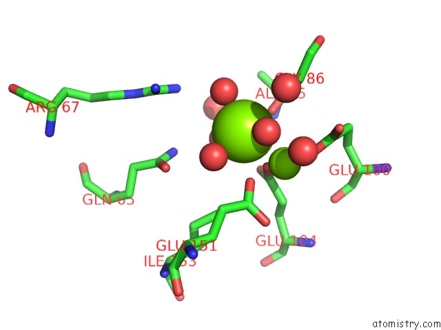
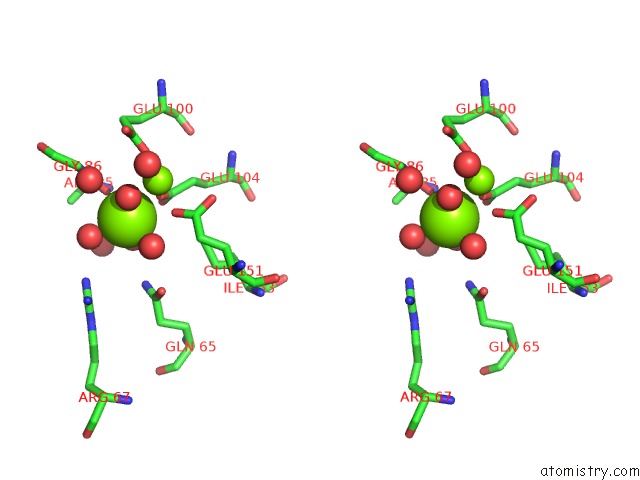
Magnesium binding site 1 out of 3 in 3o6z
Go back to
Magnesium binding site 1 out
of 3 in the Structure of the D152A E.Coli Gdp-Mannose Hydrolase (Yffh) in Complex with Mg++
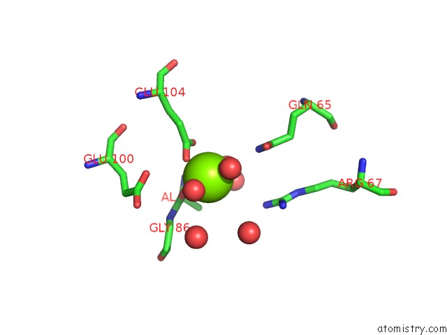
Mono view
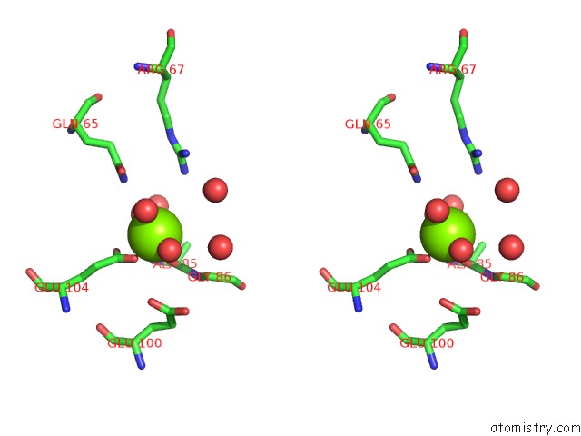
Stereo pair view
Mono view
Stereo pair view
A full contact list of Magnesium with other atoms in the Mg binding
site number 1 of Structure of the D152A E.Coli Gdp-Mannose Hydrolase (Yffh) in Complex with Mg++ within 5.0Å range:
|
Magnesium binding site 2 out of 3 in 3o6z
Go back to
Magnesium binding site 2 out
of 3 in the Structure of the D152A E.Coli Gdp-Mannose Hydrolase (Yffh) in Complex with Mg++
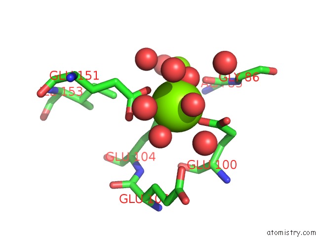
Mono view
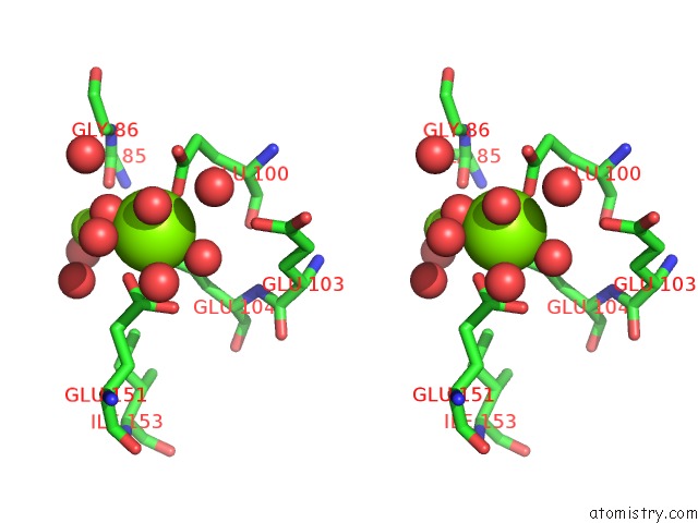
Stereo pair view
Mono view
Stereo pair view
A full contact list of Magnesium with other atoms in the Mg binding
site number 2 of Structure of the D152A E.Coli Gdp-Mannose Hydrolase (Yffh) in Complex with Mg++ within 5.0Å range:
|
Magnesium binding site 3 out of 3 in 3o6z
Go back to
Magnesium binding site 3 out
of 3 in the Structure of the D152A E.Coli Gdp-Mannose Hydrolase (Yffh) in Complex with Mg++
Mono view
Stereo pair view
Mono view
Stereo pair view
A full contact list of Magnesium with other atoms in the Mg binding
site number 3 of Structure of the D152A E.Coli Gdp-Mannose Hydrolase (Yffh) in Complex with Mg++ within 5.0Å range:
|
Reference:
A.N.Boto,
W.Xu,
J.Jakoncic,
A.Pannuri,
T.Romeo,
M.J.Bessman,
S.B.Gabelli,
L.M.Amzel.
Structural Studies of the Nudix Gdp-Mannose Hydrolase From E. Coli Reveals A New Motif For Mannose Recognition. Proteins V. 79 2455 2011.
ISSN: ISSN 0887-3585
PubMed: 21638333
DOI: 10.1002/PROT.23069
Page generated: Thu Aug 15 08:12:31 2024
ISSN: ISSN 0887-3585
PubMed: 21638333
DOI: 10.1002/PROT.23069
Last articles
K in 5KUMK in 5L9W
K in 5L9D
K in 5L88
K in 5KSD
K in 5KSE
K in 5K09
K in 5KOE
K in 5KMT
K in 5KIL