Magnesium »
PDB 3o1n-3oha »
3ocv »
Magnesium in PDB 3ocv: Structure of Recombinant Haemophilus Influenzae E(P4) Acid Phosphatase Mutant D66N Complexed with 5'-Amp
Enzymatic activity of Structure of Recombinant Haemophilus Influenzae E(P4) Acid Phosphatase Mutant D66N Complexed with 5'-Amp
All present enzymatic activity of Structure of Recombinant Haemophilus Influenzae E(P4) Acid Phosphatase Mutant D66N Complexed with 5'-Amp:
3.1.3.2;
3.1.3.2;
Protein crystallography data
The structure of Structure of Recombinant Haemophilus Influenzae E(P4) Acid Phosphatase Mutant D66N Complexed with 5'-Amp, PDB code: 3ocv
was solved by
H.Singh,
J.Schuermann,
T.Reilly,
M.Calcutt,
J.Tanner,
with X-Ray Crystallography technique. A brief refinement statistics is given in the table below:
| Resolution Low / High (Å) | 48.94 / 1.55 |
| Space group | P 65 2 2 |
| Cell size a, b, c (Å), α, β, γ (°) | 97.885, 97.885, 107.084, 90.00, 90.00, 120.00 |
| R / Rfree (%) | 14.1 / 17.1 |
Magnesium Binding Sites:
The binding sites of Magnesium atom in the Structure of Recombinant Haemophilus Influenzae E(P4) Acid Phosphatase Mutant D66N Complexed with 5'-Amp
(pdb code 3ocv). This binding sites where shown within
5.0 Angstroms radius around Magnesium atom.
In total only one binding site of Magnesium was determined in the Structure of Recombinant Haemophilus Influenzae E(P4) Acid Phosphatase Mutant D66N Complexed with 5'-Amp, PDB code: 3ocv:
In total only one binding site of Magnesium was determined in the Structure of Recombinant Haemophilus Influenzae E(P4) Acid Phosphatase Mutant D66N Complexed with 5'-Amp, PDB code: 3ocv:
Magnesium binding site 1 out of 1 in 3ocv
Go back to
Magnesium binding site 1 out
of 1 in the Structure of Recombinant Haemophilus Influenzae E(P4) Acid Phosphatase Mutant D66N Complexed with 5'-Amp
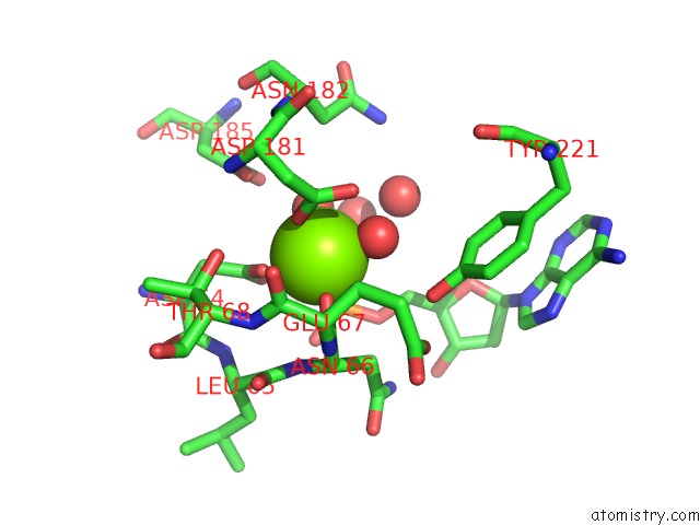
Mono view
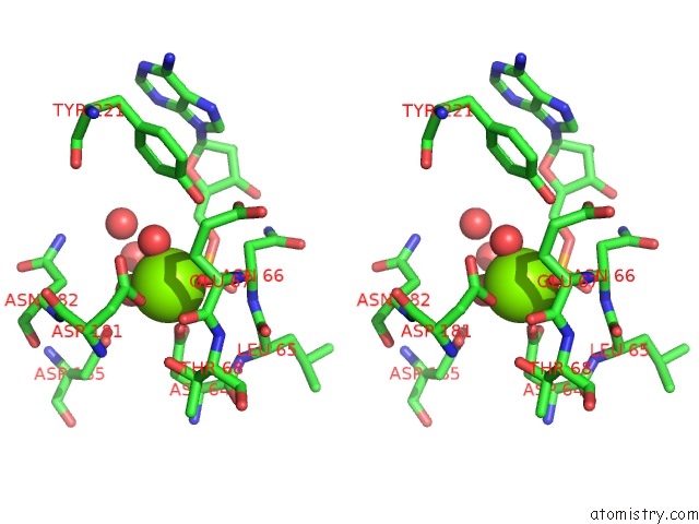
Stereo pair view
Mono view
Stereo pair view
A full contact list of Magnesium with other atoms in the Mg binding
site number 1 of Structure of Recombinant Haemophilus Influenzae E(P4) Acid Phosphatase Mutant D66N Complexed with 5'-Amp within 5.0Å range:
|
Reference:
H.Singh,
J.P.Schuermann,
T.J.Reilly,
M.J.Calcutt,
J.J.Tanner.
Recognition of Nucleoside Monophosphate Substrates By Haemophilus Influenzae Class C Acid Phosphatase. J.Mol.Biol. V. 404 639 2010.
ISSN: ISSN 0022-2836
PubMed: 20934434
DOI: 10.1016/J.JMB.2010.09.065
Page generated: Mon Aug 11 01:07:26 2025
ISSN: ISSN 0022-2836
PubMed: 20934434
DOI: 10.1016/J.JMB.2010.09.065
Last articles
Na in 6HWRNa in 6HY8
Na in 6HXI
Na in 6HUZ
Na in 6HTK
Na in 6HWP
Na in 6HUX
Na in 6HSX
Na in 6HRL
Na in 6HSN