Magnesium »
PDB 3p1g-3pio »
3pfr »
Magnesium in PDB 3pfr: Crystal Structure of D-Glucarate Dehydratase Related Protein From Actinobacillus Succinogenes Complexed with D-Glucarate
Protein crystallography data
The structure of Crystal Structure of D-Glucarate Dehydratase Related Protein From Actinobacillus Succinogenes Complexed with D-Glucarate, PDB code: 3pfr
was solved by
A.A.Fedorov,
E.V.Fedorov,
F.Mills-Groninger,
S.Ghasempur,
J.A.Gerlt,
S.C.Almo,
with X-Ray Crystallography technique. A brief refinement statistics is given in the table below:
| Resolution Low / High (Å) | 38.86 / 1.90 |
| Space group | P 41 |
| Cell size a, b, c (Å), α, β, γ (°) | 85.712, 85.712, 253.238, 90.00, 90.00, 90.00 |
| R / Rfree (%) | 21.6 / 26.3 |
Magnesium Binding Sites:
The binding sites of Magnesium atom in the Crystal Structure of D-Glucarate Dehydratase Related Protein From Actinobacillus Succinogenes Complexed with D-Glucarate
(pdb code 3pfr). This binding sites where shown within
5.0 Angstroms radius around Magnesium atom.
In total 4 binding sites of Magnesium where determined in the Crystal Structure of D-Glucarate Dehydratase Related Protein From Actinobacillus Succinogenes Complexed with D-Glucarate, PDB code: 3pfr:
Jump to Magnesium binding site number: 1; 2; 3; 4;
In total 4 binding sites of Magnesium where determined in the Crystal Structure of D-Glucarate Dehydratase Related Protein From Actinobacillus Succinogenes Complexed with D-Glucarate, PDB code: 3pfr:
Jump to Magnesium binding site number: 1; 2; 3; 4;
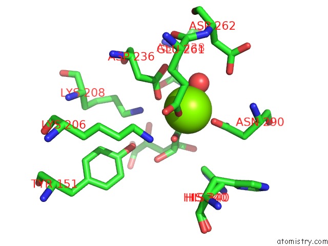
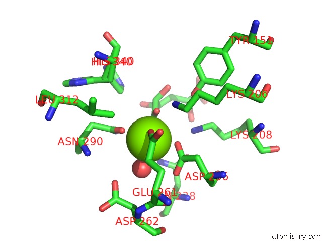
Magnesium binding site 1 out of 4 in 3pfr
Go back to
Magnesium binding site 1 out
of 4 in the Crystal Structure of D-Glucarate Dehydratase Related Protein From Actinobacillus Succinogenes Complexed with D-Glucarate
Mono view
Stereo pair view
Mono view
Stereo pair view
A full contact list of Magnesium with other atoms in the Mg binding
site number 1 of Crystal Structure of D-Glucarate Dehydratase Related Protein From Actinobacillus Succinogenes Complexed with D-Glucarate within 5.0Å range:
|
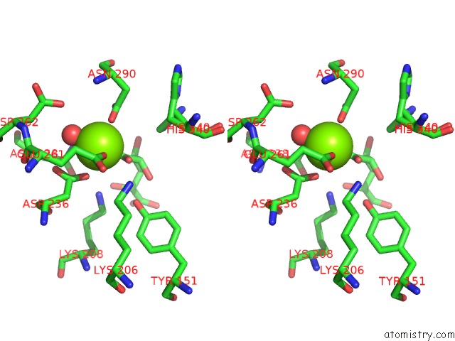
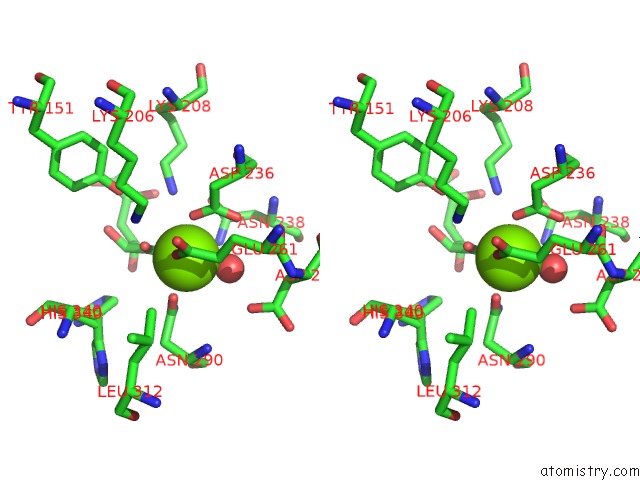
Magnesium binding site 2 out of 4 in 3pfr
Go back to
Magnesium binding site 2 out
of 4 in the Crystal Structure of D-Glucarate Dehydratase Related Protein From Actinobacillus Succinogenes Complexed with D-Glucarate
Mono view
Stereo pair view
Mono view
Stereo pair view
A full contact list of Magnesium with other atoms in the Mg binding
site number 2 of Crystal Structure of D-Glucarate Dehydratase Related Protein From Actinobacillus Succinogenes Complexed with D-Glucarate within 5.0Å range:
|
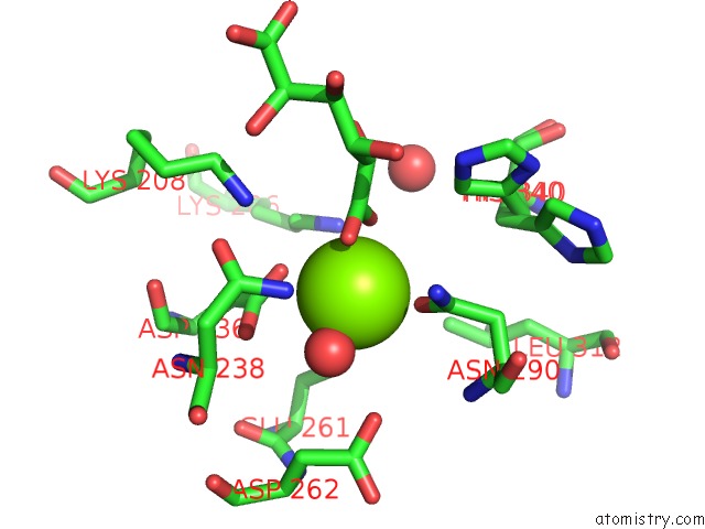
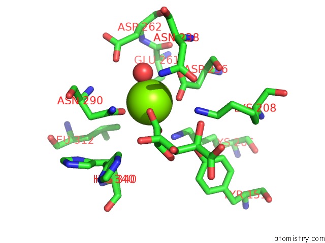
Magnesium binding site 3 out of 4 in 3pfr
Go back to
Magnesium binding site 3 out
of 4 in the Crystal Structure of D-Glucarate Dehydratase Related Protein From Actinobacillus Succinogenes Complexed with D-Glucarate
Mono view
Stereo pair view
Mono view
Stereo pair view
A full contact list of Magnesium with other atoms in the Mg binding
site number 3 of Crystal Structure of D-Glucarate Dehydratase Related Protein From Actinobacillus Succinogenes Complexed with D-Glucarate within 5.0Å range:
|
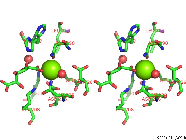
Magnesium binding site 4 out of 4 in 3pfr
Go back to
Magnesium binding site 4 out
of 4 in the Crystal Structure of D-Glucarate Dehydratase Related Protein From Actinobacillus Succinogenes Complexed with D-Glucarate
Mono view
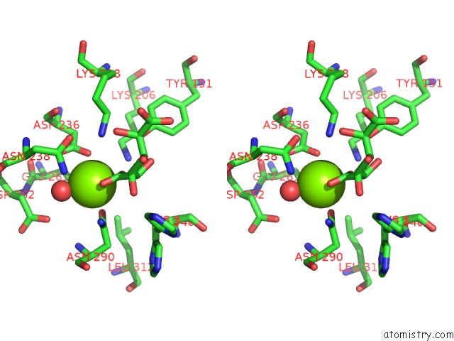
Stereo pair view
Mono view
Stereo pair view
A full contact list of Magnesium with other atoms in the Mg binding
site number 4 of Crystal Structure of D-Glucarate Dehydratase Related Protein From Actinobacillus Succinogenes Complexed with D-Glucarate within 5.0Å range:
|
Reference:
A.A.Fedorov,
E.V.Fedorov,
F.Mills-Groninger,
S.Ghasempur,
J.A.Gerlt,
S.C.Almo.
Crystal Structure of D-Glucarate Dehydratase Related Protein From Actinobacillus Succinogenes Complexed with D-Glucarate To Be Published.
Page generated: Mon Aug 11 01:50:39 2025
Last articles
Mg in 7PLHMg in 7PJI
Mg in 7PJE
Mg in 7PJF
Mg in 7PH4
Mg in 7PDZ
Mg in 7PH3
Mg in 7PGI
Mg in 7PEM
Mg in 7PDS