Atomistry » Magnesium » PDB 3pip-3px4 » 3pk7
Atomistry »
  Magnesium »
    PDB 3pip-3px4 »
      3pk7 »

Magnesium in PDB 3pk7: Crystal Structure of D-Mannonate Dehydratase From Chromohalobacter Salexigens with Mg and Glycerol Bound in the Active Site

Protein crystallography data

The structure of Crystal Structure of D-Mannonate Dehydratase From Chromohalobacter Salexigens with Mg and Glycerol Bound in the Active Site, PDB code: 3pk7 was solved by A.A.Fedorov, E.V.Fedorov, D.Wichelecki, J.A.Gerlt, S.C.Almo, with X-Ray Crystallography technique. A brief refinement statistics is given in the table below:

Resolution Low / High (Å) 40.05 / 1.64
Space group C 1 2 1
Cell size a, b, c (Å), α, β, γ (°) 195.273, 85.762, 195.097, 90.00, 110.31, 90.00
R / Rfree (%) 15.8 / 18.4

Magnesium Binding Sites:

The binding sites of Magnesium atom in the Crystal Structure of D-Mannonate Dehydratase From Chromohalobacter Salexigens with Mg and Glycerol Bound in the Active Site (pdb code 3pk7). This binding sites where shown within 5.0 Angstroms radius around Magnesium atom.
In total 9 binding sites of Magnesium where determined in the Crystal Structure of D-Mannonate Dehydratase From Chromohalobacter Salexigens with Mg and Glycerol Bound in the Active Site, PDB code: 3pk7:
Jump to Magnesium binding site number: 1; 2; 3; 4; 5; 6; 7; 8; 9;

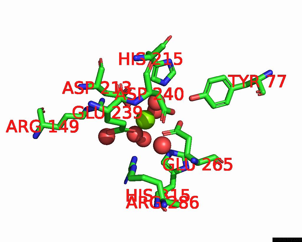
Magnesium binding site 1 out of 9 in 3pk7

Go back to Magnesium Binding Sites List in 3pk7
Magnesium binding site 1 out of 9 in the Crystal Structure of D-Mannonate Dehydratase From Chromohalobacter Salexigens with Mg and Glycerol Bound in the Active Site


Mono view


Stereo pair view

A full contact list of Magnesium with other atoms in the Mg binding site number 1 of Crystal Structure of D-Mannonate Dehydratase From Chromohalobacter Salexigens with Mg and Glycerol Bound in the Active Site within 5.0Å range:
probe atom residue distance (Å) B Occ
A:Mg406

b:16.2
occ:1.00
OD2 A:ASP213 2.1 14.8 1.0
OE1 A:GLU239 2.1 15.4 1.0
OE1 A:GLU265 2.1 15.7 1.0
O A:HOH458 2.2 17.1 1.0
O A:HOH423 2.2 15.6 1.0
O A:HOH409 2.2 14.2 1.0
CD A:GLU265 3.0 13.7 1.0
CG A:ASP213 3.1 16.9 1.0
CD A:GLU239 3.1 16.1 1.0
OE2 A:GLU265 3.3 15.5 1.0
OD1 A:ASP213 3.5 14.5 1.0
NH2 A:ARG286 3.7 16.5 1.0
OE2 A:GLU239 3.8 14.1 1.0
O A:HOH418 4.0 15.9 1.0
CD2 A:HIS215 4.0 18.5 1.0
CG A:GLU239 4.1 14.1 1.0
O A:HOH454 4.2 18.9 1.0
OD2 A:ASP240 4.2 15.2 1.0
O A:HOH435 4.3 14.5 1.0
CB A:ASP213 4.3 16.1 1.0
CG A:GLU265 4.4 13.8 1.0
NH1 A:ARG149 4.4 20.7 1.0
NE2 A:HIS215 4.4 21.2 1.0
OH A:TYR161 4.4 28.8 1.0
OH B:TYR77 4.7 13.9 1.0
CG A:ASP240 4.8 14.9 1.0
CZ A:ARG286 4.8 18.5 1.0
NE2 A:HIS315 4.8 19.1 1.0
CD2 A:HIS315 4.8 16.1 1.0
NE A:ARG286 5.0 20.1 1.0

Magnesium binding site 2 out of 9 in 3pk7

Go back to Magnesium Binding Sites List in 3pk7
Magnesium binding site 2 out of 9 in the Crystal Structure of D-Mannonate Dehydratase From Chromohalobacter Salexigens with Mg and Glycerol Bound in the Active Site


Mono view


Stereo pair view

A full contact list of Magnesium with other atoms in the Mg binding site number 2 of Crystal Structure of D-Mannonate Dehydratase From Chromohalobacter Salexigens with Mg and Glycerol Bound in the Active Site within 5.0Å range:
probe atom residue distance (Å) B Occ
B:Mg406

b:14.4
occ:1.00
OD2 B:ASP213 2.1 11.8 1.0
OE1 B:GLU239 2.1 13.1 1.0
O B:HOH411 2.2 13.8 1.0
OE1 B:GLU265 2.2 13.1 1.0
O B:HOH455 2.2 15.4 1.0
O B:HOH426 2.2 14.7 1.0
CD B:GLU265 3.1 14.2 1.0
CG B:ASP213 3.1 11.8 1.0
CD B:GLU239 3.2 13.1 1.0
OE2 B:GLU265 3.3 15.6 1.0
OD1 B:ASP213 3.5 13.7 1.0
NH2 B:ARG286 3.8 16.6 1.0
OE2 B:GLU239 3.9 12.7 1.0
O B:HOH432 4.0 14.5 1.0
CD2 B:HIS215 4.0 23.8 1.0
CG B:GLU239 4.1 12.8 1.0
OD2 B:ASP240 4.1 15.1 1.0
O B:HOH445 4.2 14.2 1.0
O B:HOH434 4.3 17.9 1.0
NH1 B:ARG149 4.3 21.2 1.0
NE2 B:HIS215 4.3 26.9 1.0
CB B:ASP213 4.3 13.9 1.0
CG B:GLU265 4.4 12.1 1.0
CG B:ASP240 4.7 14.7 1.0
OH A:TYR77 4.7 16.1 1.0
CZ B:ARG286 4.8 18.7 1.0
NE2 B:HIS315 4.9 17.4 1.0
CD2 B:HIS315 4.9 14.4 1.0
NE B:ARG286 4.9 19.8 1.0

Magnesium binding site 3 out of 9 in 3pk7

Go back to Magnesium Binding Sites List in 3pk7
Magnesium binding site 3 out of 9 in the Crystal Structure of D-Mannonate Dehydratase From Chromohalobacter Salexigens with Mg and Glycerol Bound in the Active Site


Mono view


Stereo pair view

A full contact list of Magnesium with other atoms in the Mg binding site number 3 of Crystal Structure of D-Mannonate Dehydratase From Chromohalobacter Salexigens with Mg and Glycerol Bound in the Active Site within 5.0Å range:
probe atom residue distance (Å) B Occ
C:Mg406

b:13.8
occ:1.00
OD2 C:ASP213 2.1 13.0 1.0
OE1 C:GLU239 2.1 12.4 1.0
OE1 C:GLU265 2.1 13.6 1.0
O C:HOH408 2.2 13.9 1.0
O C:HOH445 2.2 15.6 1.0
O C:HOH442 2.2 15.5 1.0
CD C:GLU265 3.0 15.2 1.0
CG C:ASP213 3.1 13.9 1.0
CD C:GLU239 3.1 14.3 1.0
OE2 C:GLU265 3.3 14.6 1.0
OD1 C:ASP213 3.5 12.6 1.0
NH2 C:ARG286 3.8 15.7 1.0
OE2 C:GLU239 3.8 12.7 1.0
O C:HOH456 4.0 15.7 1.0
CD2 C:HIS215 4.0 18.9 1.0
CG C:GLU239 4.1 13.1 1.0
OD2 C:ASP240 4.1 15.4 1.0
O C:HOH416 4.3 13.7 1.0
O C:HOH460 4.3 18.0 1.0
CB C:ASP213 4.4 14.4 1.0
CG C:GLU265 4.4 14.3 1.0
NH1 C:ARG149 4.4 18.4 1.0
NE2 C:HIS215 4.4 22.6 1.0
OH C:TYR161 4.4 28.1 1.0
CG C:ASP240 4.7 15.9 1.0
OH D:TYR77 4.8 14.0 1.0
CZ C:ARG286 4.9 17.5 1.0
CD2 C:HIS315 4.9 16.8 1.0
NE2 C:HIS315 4.9 17.4 1.0
NE C:ARG286 5.0 22.3 1.0

Magnesium binding site 4 out of 9 in 3pk7

Go back to Magnesium Binding Sites List in 3pk7
Magnesium binding site 4 out of 9 in the Crystal Structure of D-Mannonate Dehydratase From Chromohalobacter Salexigens with Mg and Glycerol Bound in the Active Site


Mono view


Stereo pair view

A full contact list of Magnesium with other atoms in the Mg binding site number 4 of Crystal Structure of D-Mannonate Dehydratase From Chromohalobacter Salexigens with Mg and Glycerol Bound in the Active Site within 5.0Å range:
probe atom residue distance (Å) B Occ
D:Mg406

b:17.6
occ:1.00
OE1 D:GLU239 2.1 16.0 1.0
OD2 D:ASP213 2.1 14.8 1.0
OE1 D:GLU265 2.1 16.4 1.0
O D:HOH942 2.1 16.4 1.0
O D:HOH441 2.3 17.5 1.0
O D:HOH413 2.3 15.6 1.0
CD D:GLU265 3.0 16.5 1.0
CG D:ASP213 3.1 15.3 1.0
CD D:GLU239 3.1 19.1 1.0
OE2 D:GLU265 3.3 18.4 1.0
OD1 D:ASP213 3.5 15.8 1.0
NH2 D:ARG286 3.7 18.4 1.0
OE2 D:GLU239 3.8 17.0 1.0
CD2 D:HIS215 3.9 23.9 1.0
O D:HOH416 4.0 17.8 1.0
CG D:GLU239 4.1 14.6 1.0
OD2 D:ASP240 4.1 16.6 1.0
NE2 D:HIS215 4.2 29.5 1.0
O D:HOH435 4.2 21.1 1.0
O D:HOH409 4.3 15.8 1.0
CB D:ASP213 4.4 16.7 1.0
NH1 D:ARG149 4.4 25.8 1.0
CG D:GLU265 4.4 15.0 1.0
OH C:TYR77 4.7 15.6 1.0
CG D:ASP240 4.7 17.1 1.0
CZ D:ARG286 4.8 18.9 1.0
NE2 D:HIS315 4.8 19.6 1.0
CD2 D:HIS315 4.9 18.1 1.0
NE D:ARG286 4.9 23.3 1.0

Magnesium binding site 5 out of 9 in 3pk7

Go back to Magnesium Binding Sites List in 3pk7
Magnesium binding site 5 out of 9 in the Crystal Structure of D-Mannonate Dehydratase From Chromohalobacter Salexigens with Mg and Glycerol Bound in the Active Site


Mono view


Stereo pair view

A full contact list of Magnesium with other atoms in the Mg binding site number 5 of Crystal Structure of D-Mannonate Dehydratase From Chromohalobacter Salexigens with Mg and Glycerol Bound in the Active Site within 5.0Å range:
probe atom residue distance (Å) B Occ
D:Mg408

b:40.6
occ:1.00
O D:HOH2745 2.4 36.1 1.0
O D:HOH3071 2.4 36.5 1.0
O D:HOH1862 2.4 30.0 1.0
O D:HOH3073 2.5 41.2 1.0
O D:HOH3072 2.5 37.5 1.0
O D:HOH2559 2.5 38.1 1.0
OD1 D:ASP376 4.2 29.6 1.0
O D:PRO371 4.2 20.9 1.0
OD2 D:ASP376 4.2 39.1 1.0
O D:HOH2569 4.3 34.7 1.0
OG D:SER370 4.4 30.3 1.0
O D:HOH756 4.4 23.2 1.0
O D:HOH2065 4.6 32.8 1.0
CG D:ASP376 4.6 33.1 1.0
CE D:LYS101 4.7 19.3 1.0
CB D:SER370 4.7 25.0 1.0
NZ D:LYS101 5.0 18.4 1.0

Magnesium binding site 6 out of 9 in 3pk7

Go back to Magnesium Binding Sites List in 3pk7
Magnesium binding site 6 out of 9 in the Crystal Structure of D-Mannonate Dehydratase From Chromohalobacter Salexigens with Mg and Glycerol Bound in the Active Site


Mono view


Stereo pair view

A full contact list of Magnesium with other atoms in the Mg binding site number 6 of Crystal Structure of D-Mannonate Dehydratase From Chromohalobacter Salexigens with Mg and Glycerol Bound in the Active Site within 5.0Å range:
probe atom residue distance (Å) B Occ
E:Mg406

b:14.8
occ:1.00
OE1 E:GLU239 2.1 13.4 1.0
OE1 E:GLU265 2.1 12.4 1.0
OD2 E:ASP213 2.1 13.6 1.0
O E:HOH416 2.1 15.0 1.0
O E:HOH437 2.2 15.8 1.0
O E:HOH428 2.2 16.4 1.0
CD E:GLU265 3.0 15.0 1.0
CG E:ASP213 3.1 16.1 1.0
CD E:GLU239 3.1 14.6 1.0
OE2 E:GLU265 3.3 16.1 1.0
OD1 E:ASP213 3.5 14.5 1.0
NH2 E:ARG286 3.8 14.5 1.0
OE2 E:GLU239 3.9 13.4 1.0
CD2 E:HIS215 4.0 21.3 1.0
O E:HOH418 4.0 14.8 1.0
CG E:GLU239 4.1 13.2 1.0
OD2 E:ASP240 4.1 15.2 1.0
O E:HOH413 4.2 14.7 1.0
O E:HOH447 4.3 17.7 1.0
NH1 E:ARG149 4.3 19.4 1.0
NE2 E:HIS215 4.3 22.1 1.0
CG E:GLU265 4.4 14.5 1.0
CB E:ASP213 4.4 15.5 1.0
OH E:TYR161 4.5 29.2 1.0
CG E:ASP240 4.7 14.3 1.0
OH H:TYR77 4.7 16.4 1.0
CZ E:ARG286 4.8 19.4 1.0
NE2 E:HIS315 4.9 16.9 1.0
CD2 E:HIS315 4.9 13.3 1.0
NE E:ARG286 5.0 20.9 1.0
CB E:GLU265 5.0 15.0 1.0

Magnesium binding site 7 out of 9 in 3pk7

Go back to Magnesium Binding Sites List in 3pk7
Magnesium binding site 7 out of 9 in the Crystal Structure of D-Mannonate Dehydratase From Chromohalobacter Salexigens with Mg and Glycerol Bound in the Active Site


Mono view


Stereo pair view

A full contact list of Magnesium with other atoms in the Mg binding site number 7 of Crystal Structure of D-Mannonate Dehydratase From Chromohalobacter Salexigens with Mg and Glycerol Bound in the Active Site within 5.0Å range:
probe atom residue distance (Å) B Occ
F:Mg406

b:15.1
occ:1.00
OD2 F:ASP213 2.1 14.3 1.0
OE1 F:GLU239 2.1 14.4 1.0
OE1 F:GLU265 2.1 14.2 1.0
O F:HOH420 2.2 14.6 1.0
O F:HOH458 2.2 16.9 1.0
O F:HOH414 2.2 15.7 1.0
CD F:GLU265 3.0 14.8 1.0
CG F:ASP213 3.1 14.3 1.0
CD F:GLU239 3.1 11.5 1.0
OE2 F:GLU265 3.3 18.2 1.0
OD1 F:ASP213 3.4 14.2 1.0
NH2 F:ARG286 3.8 14.0 1.0
OE2 F:GLU239 3.9 15.9 1.0
O F:HOH408 3.9 16.6 1.0
CD2 F:HIS215 4.0 26.3 1.0
CG F:GLU239 4.1 13.9 1.0
OD2 F:ASP240 4.2 14.9 1.0
NH1 F:ARG149 4.3 22.0 1.0
NE2 F:HIS215 4.3 27.0 1.0
O F:HOH415 4.3 13.3 1.0
CB F:ASP213 4.3 14.8 1.0
O F:HOH452 4.3 19.4 1.0
CG F:GLU265 4.4 15.7 1.0
OH G:TYR77 4.7 13.9 1.0
CG F:ASP240 4.8 15.3 1.0
CZ F:ARG286 4.8 17.0 1.0
NE2 F:HIS315 4.9 18.5 1.0
CD2 F:HIS315 4.9 13.3 1.0
NE F:ARG286 4.9 20.9 1.0

Magnesium binding site 8 out of 9 in 3pk7

Go back to Magnesium Binding Sites List in 3pk7
Magnesium binding site 8 out of 9 in the Crystal Structure of D-Mannonate Dehydratase From Chromohalobacter Salexigens with Mg and Glycerol Bound in the Active Site


Mono view


Stereo pair view

A full contact list of Magnesium with other atoms in the Mg binding site number 8 of Crystal Structure of D-Mannonate Dehydratase From Chromohalobacter Salexigens with Mg and Glycerol Bound in the Active Site within 5.0Å range:
probe atom residue distance (Å) B Occ
G:Mg406

b:14.1
occ:1.00
OD2 G:ASP213 2.1 14.0 1.0
OE1 G:GLU239 2.1 14.7 1.0
OE1 G:GLU265 2.1 13.4 1.0
O G:HOH414 2.2 14.5 1.0
O G:HOH441 2.2 16.3 1.0
O G:HOH421 2.2 16.6 1.0
CD G:GLU265 3.0 13.1 1.0
CG G:ASP213 3.1 17.4 1.0
CD G:GLU239 3.1 14.8 1.0
OE2 G:GLU265 3.3 15.9 1.0
OD1 G:ASP213 3.4 13.9 1.0
NH2 G:ARG286 3.8 15.9 1.0
OE2 G:GLU239 3.8 14.8 1.0
O G:HOH412 4.0 15.4 1.0
CD2 G:HIS215 4.0 24.8 1.0
CG G:GLU239 4.0 12.1 1.0
OD2 G:ASP240 4.2 14.7 1.0
O G:HOH415 4.2 13.0 1.0
NH1 G:ARG149 4.3 21.8 1.0
NE2 G:HIS215 4.3 24.1 1.0
CG G:GLU265 4.3 11.7 1.0
O G:HOH3041 4.4 19.0 1.0
CB G:ASP213 4.4 13.6 1.0
OH G:TYR161 4.4 31.3 1.0
OH F:TYR77 4.7 13.8 1.0
CG G:ASP240 4.7 13.5 1.0
NE2 G:HIS315 4.8 17.9 1.0
CZ G:ARG286 4.8 19.8 1.0
CD2 G:HIS315 4.9 15.7 1.0
NE G:ARG286 5.0 21.5 1.0
CZ G:TYR161 5.0 29.1 1.0

Magnesium binding site 9 out of 9 in 3pk7

Go back to Magnesium Binding Sites List in 3pk7
Magnesium binding site 9 out of 9 in the Crystal Structure of D-Mannonate Dehydratase From Chromohalobacter Salexigens with Mg and Glycerol Bound in the Active Site


Mono view


Stereo pair view

A full contact list of Magnesium with other atoms in the Mg binding site number 9 of Crystal Structure of D-Mannonate Dehydratase From Chromohalobacter Salexigens with Mg and Glycerol Bound in the Active Site within 5.0Å range:
probe atom residue distance (Å) B Occ
H:Mg406

b:17.0
occ:1.00
OE1 H:GLU239 2.1 15.5 1.0
OD2 H:ASP213 2.1 17.8 1.0
O H:HOH416 2.1 16.2 1.0
OE1 H:GLU265 2.1 14.6 1.0
O H:HOH418 2.2 17.9 1.0
O H:HOH574 2.3 22.3 1.0
CD H:GLU265 3.0 14.9 1.0
CG H:ASP213 3.1 17.0 1.0
CD H:GLU239 3.1 15.6 1.0
OE2 H:GLU265 3.2 18.1 1.0
OD1 H:ASP213 3.5 16.4 1.0
NH2 H:ARG286 3.8 17.3 1.0
OE2 H:GLU239 3.8 18.1 1.0
CD2 H:HIS215 3.9 22.7 1.0
O H:HOH420 4.0 17.4 1.0
CG H:GLU239 4.1 15.0 1.0
OD2 H:ASP240 4.1 17.7 1.0
O H:HOH415 4.3 15.9 1.0
NE2 H:HIS215 4.3 26.1 1.0
CG H:GLU265 4.3 17.2 1.0
O H:HOH932 4.3 21.5 1.0
CB H:ASP213 4.4 15.9 1.0
NH1 H:ARG149 4.4 25.4 1.0
OH H:TYR161 4.4 35.9 1.0
OH E:TYR77 4.7 16.1 1.0
CG H:ASP240 4.7 16.3 1.0
CZ H:ARG286 4.8 19.0 1.0
CD2 H:HIS315 4.9 19.7 1.0
NE2 H:HIS315 4.9 20.8 1.0
NE H:ARG286 4.9 24.0 1.0
CB H:GLU265 5.0 15.0 1.0

Reference:

A.A.Fedorov, E.V.Fedorov, D.Wichelecki, J.A.Gerlt, S.C.Almo. Crystal Structure of D-Mannonate Dehydratase From Chromohalobacter Salexigens with Mg and Glycerol Bound in the Active Site To Be Published.
Page generated: Thu Aug 15 09:35:31 2024

Last articles

Fe in 2YXO
Fe in 2YRS
Fe in 2YXC
Fe in 2YNM
Fe in 2YVJ
Fe in 2YP1
Fe in 2YU2
Fe in 2YU1
Fe in 2YQB
Fe in 2YOO
© Copyright 2008-2020 by atomistry.com
Home   |    Site Map   |    Copyright   |    Contact us   |    Privacy