Magnesium »
PDB 3r11-3ren »
3r4v »
Magnesium in PDB 3r4v: Structure of the Phage Tubulin Phuz-Gdp
Protein crystallography data
The structure of Structure of the Phage Tubulin Phuz-Gdp, PDB code: 3r4v
was solved by
D.A.Agard,
J.Pogliano,
J.A.Kraemer,
M.L.Erb,
C.A.Waddling,
E.A.Montabana,
H.Wang,
K.Nguyen,
S.Pham,
with X-Ray Crystallography technique. A brief refinement statistics is given in the table below:
| Resolution Low / High (Å) | 46.40 / 1.67 |
| Space group | P 21 21 21 |
| Cell size a, b, c (Å), α, β, γ (°) | 47.065, 75.935, 92.798, 90.00, 90.00, 90.00 |
| R / Rfree (%) | 18.6 / 23.2 |
Magnesium Binding Sites:
The binding sites of Magnesium atom in the Structure of the Phage Tubulin Phuz-Gdp
(pdb code 3r4v). This binding sites where shown within
5.0 Angstroms radius around Magnesium atom.
In total only one binding site of Magnesium was determined in the Structure of the Phage Tubulin Phuz-Gdp, PDB code: 3r4v:
In total only one binding site of Magnesium was determined in the Structure of the Phage Tubulin Phuz-Gdp, PDB code: 3r4v:
Magnesium binding site 1 out of 1 in 3r4v
Go back to
Magnesium binding site 1 out
of 1 in the Structure of the Phage Tubulin Phuz-Gdp
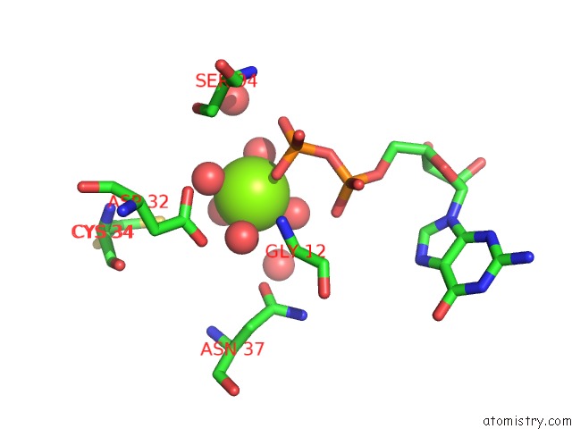
Mono view
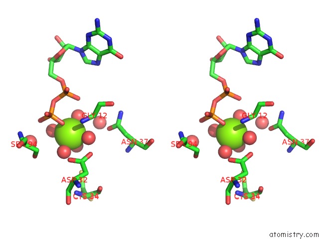
Stereo pair view
Mono view
Stereo pair view
A full contact list of Magnesium with other atoms in the Mg binding
site number 1 of Structure of the Phage Tubulin Phuz-Gdp within 5.0Å range:
|
Reference:
J.A.Kraemer,
M.L.Erb,
C.A.Waddling,
E.A.Montabana,
E.A.Zehr,
H.Wang,
K.Nguyen,
D.S.Pham,
D.A.Agard,
J.Pogliano.
A Phage Tubulin Assembles Dynamic Filaments By An Atypical Mechanism to Center Viral Dna Within the Host Cell. Cell(Cambridge,Mass.) V. 149 1488 2012.
ISSN: ISSN 0092-8674
PubMed: 22726436
DOI: 10.1016/J.CELL.2012.04.034
Page generated: Mon Aug 11 02:35:32 2025
ISSN: ISSN 0092-8674
PubMed: 22726436
DOI: 10.1016/J.CELL.2012.04.034
Last articles
Mg in 3T2CMg in 3T2B
Mg in 3T1R
Mg in 3T1O
Mg in 3T0D
Mg in 3T1Q
Mg in 3T12
Mg in 3T1K
Mg in 3T10
Mg in 3T0Z