Magnesium »
PDB 3rr7-3rv6 »
3rv6 »
Magnesium in PDB 3rv6: Structure of A M. Tuberculosis Salicylate Synthase, Mbti, in Complex with An Inhibitor with Phenyl R-Group
Enzymatic activity of Structure of A M. Tuberculosis Salicylate Synthase, Mbti, in Complex with An Inhibitor with Phenyl R-Group
All present enzymatic activity of Structure of A M. Tuberculosis Salicylate Synthase, Mbti, in Complex with An Inhibitor with Phenyl R-Group:
5.4.4.2;
5.4.4.2;
Protein crystallography data
The structure of Structure of A M. Tuberculosis Salicylate Synthase, Mbti, in Complex with An Inhibitor with Phenyl R-Group, PDB code: 3rv6
was solved by
G.Chi,
E.M.M.Bulloch,
A.Manos-Turvey,
R.J.Payne,
J.S.Lott,
Tb Structuralgenomics Consortium (Tbsgc),
with X-Ray Crystallography technique. A brief refinement statistics is given in the table below:
| Resolution Low / High (Å) | 16.72 / 2.04 |
| Space group | P 1 21 1 |
| Cell size a, b, c (Å), α, β, γ (°) | 52.748, 91.530, 96.466, 90.00, 104.83, 90.00 |
| R / Rfree (%) | 17.1 / 20.5 |
Magnesium Binding Sites:
The binding sites of Magnesium atom in the Structure of A M. Tuberculosis Salicylate Synthase, Mbti, in Complex with An Inhibitor with Phenyl R-Group
(pdb code 3rv6). This binding sites where shown within
5.0 Angstroms radius around Magnesium atom.
In total 2 binding sites of Magnesium where determined in the Structure of A M. Tuberculosis Salicylate Synthase, Mbti, in Complex with An Inhibitor with Phenyl R-Group, PDB code: 3rv6:
Jump to Magnesium binding site number: 1; 2;
In total 2 binding sites of Magnesium where determined in the Structure of A M. Tuberculosis Salicylate Synthase, Mbti, in Complex with An Inhibitor with Phenyl R-Group, PDB code: 3rv6:
Jump to Magnesium binding site number: 1; 2;
Magnesium binding site 1 out of 2 in 3rv6
Go back to
Magnesium binding site 1 out
of 2 in the Structure of A M. Tuberculosis Salicylate Synthase, Mbti, in Complex with An Inhibitor with Phenyl R-Group
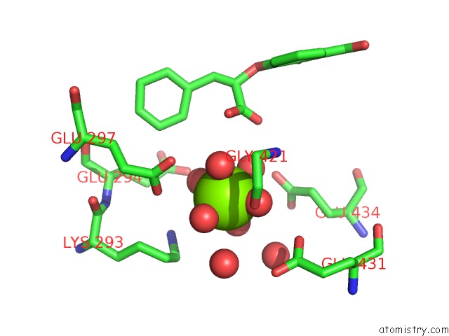
Mono view
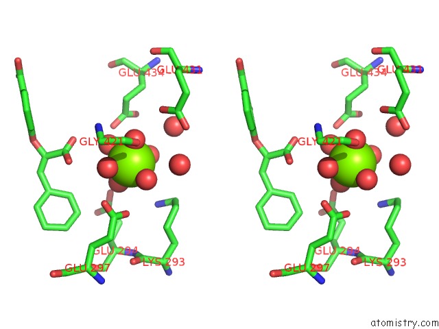
Stereo pair view
Mono view
Stereo pair view
A full contact list of Magnesium with other atoms in the Mg binding
site number 1 of Structure of A M. Tuberculosis Salicylate Synthase, Mbti, in Complex with An Inhibitor with Phenyl R-Group within 5.0Å range:
|
Magnesium binding site 2 out of 2 in 3rv6
Go back to
Magnesium binding site 2 out
of 2 in the Structure of A M. Tuberculosis Salicylate Synthase, Mbti, in Complex with An Inhibitor with Phenyl R-Group
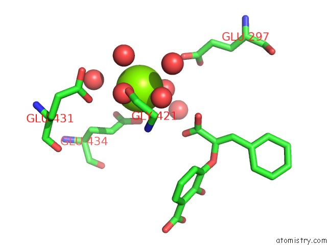
Mono view
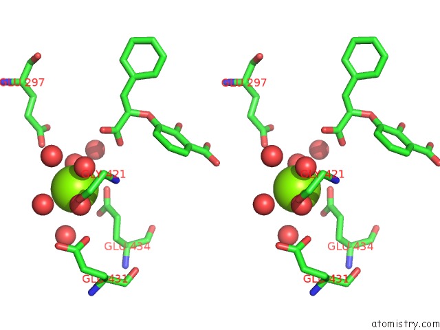
Stereo pair view
Mono view
Stereo pair view
A full contact list of Magnesium with other atoms in the Mg binding
site number 2 of Structure of A M. Tuberculosis Salicylate Synthase, Mbti, in Complex with An Inhibitor with Phenyl R-Group within 5.0Å range:
|
Reference:
G.Chi,
A.Manos-Turvey,
P.D.O'connor,
J.M.Johnston,
G.L.Evans,
E.N.Baker,
R.J.Payne,
J.S.Lott,
E.M.Bulloch.
Implications of Binding Mode and Active Site Flexibility For Inhibitor Potency Against the Salicylate Synthase From Mycobacterium Tuberculosis Biochemistry V. 51 4868 2012.
ISSN: ISSN 0006-2960
PubMed: 22607697
DOI: 10.1021/BI3002067
Page generated: Mon Aug 11 02:52:38 2025
ISSN: ISSN 0006-2960
PubMed: 22607697
DOI: 10.1021/BI3002067
Last articles
Mg in 4DUXMg in 4DUW
Mg in 4DUV
Mg in 4DUO
Mg in 4DUG
Mg in 4DTY
Mg in 4DTW
Mg in 4DTH
Mg in 4DTF
Mg in 4DSU