Magnesium »
PDB 3s99-3si8 »
3shz »
Magnesium in PDB 3shz: Crystal Structure of the PDE5A1 Catalytic Domain in Complex with Novel Inhibitors
Enzymatic activity of Crystal Structure of the PDE5A1 Catalytic Domain in Complex with Novel Inhibitors
All present enzymatic activity of Crystal Structure of the PDE5A1 Catalytic Domain in Complex with Novel Inhibitors:
3.1.4.35;
3.1.4.35;
Protein crystallography data
The structure of Crystal Structure of the PDE5A1 Catalytic Domain in Complex with Novel Inhibitors, PDB code: 3shz
was solved by
T.T.Chen,
T.Chen,
Y.C.Xu,
with X-Ray Crystallography technique. A brief refinement statistics is given in the table below:
| Resolution Low / High (Å) | 46.15 / 2.45 |
| Space group | P 31 2 1 |
| Cell size a, b, c (Å), α, β, γ (°) | 74.687, 74.687, 131.728, 90.00, 90.00, 120.00 |
| R / Rfree (%) | 20.1 / 24.5 |
Other elements in 3shz:
The structure of Crystal Structure of the PDE5A1 Catalytic Domain in Complex with Novel Inhibitors also contains other interesting chemical elements:
| Chlorine | (Cl) | 1 atom |
| Zinc | (Zn) | 1 atom |
Magnesium Binding Sites:
The binding sites of Magnesium atom in the Crystal Structure of the PDE5A1 Catalytic Domain in Complex with Novel Inhibitors
(pdb code 3shz). This binding sites where shown within
5.0 Angstroms radius around Magnesium atom.
In total 5 binding sites of Magnesium where determined in the Crystal Structure of the PDE5A1 Catalytic Domain in Complex with Novel Inhibitors, PDB code: 3shz:
Jump to Magnesium binding site number: 1; 2; 3; 4; 5;
In total 5 binding sites of Magnesium where determined in the Crystal Structure of the PDE5A1 Catalytic Domain in Complex with Novel Inhibitors, PDB code: 3shz:
Jump to Magnesium binding site number: 1; 2; 3; 4; 5;
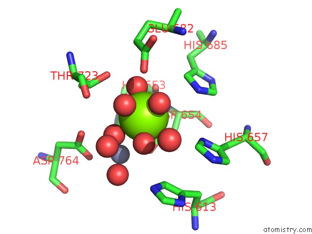
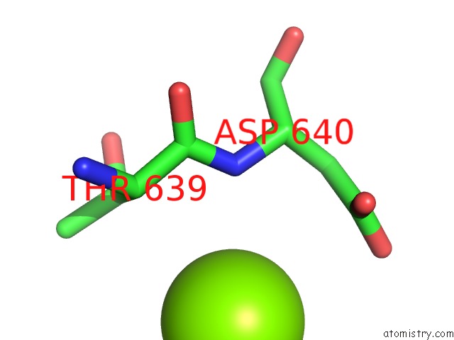
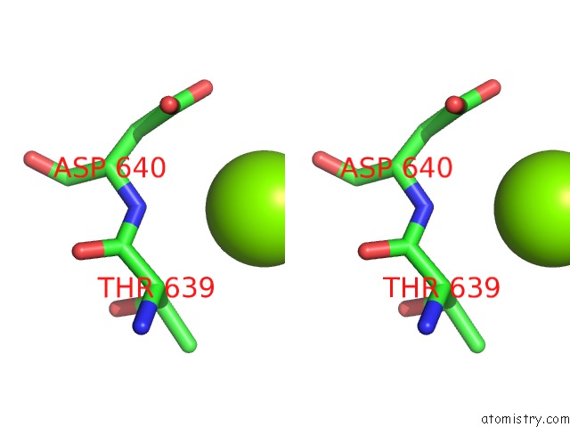
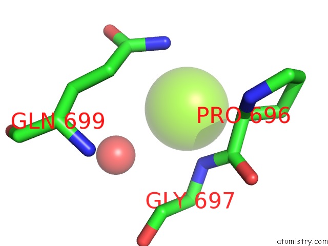
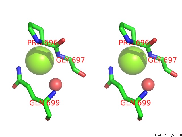
Magnesium binding site 1 out of 5 in 3shz
Go back to
Magnesium binding site 1 out
of 5 in the Crystal Structure of the PDE5A1 Catalytic Domain in Complex with Novel Inhibitors
Mono view
Stereo pair view
Mono view
Stereo pair view
A full contact list of Magnesium with other atoms in the Mg binding
site number 1 of Crystal Structure of the PDE5A1 Catalytic Domain in Complex with Novel Inhibitors within 5.0Å range:
|
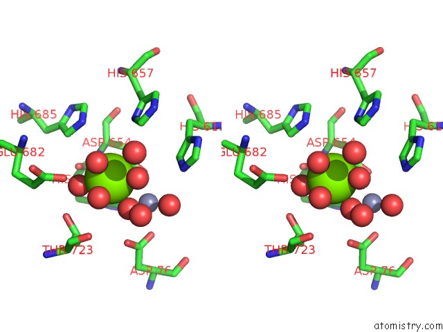
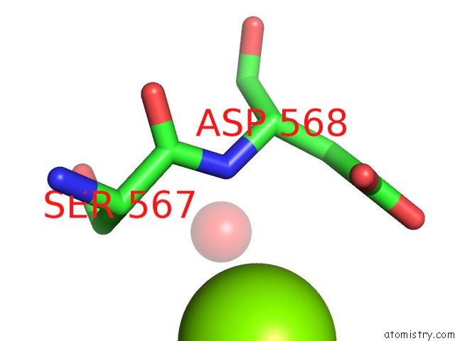
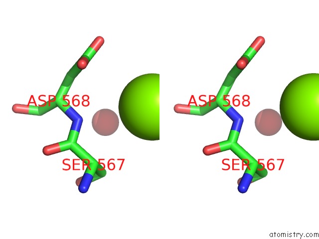
Magnesium binding site 2 out of 5 in 3shz
Go back to
Magnesium binding site 2 out
of 5 in the Crystal Structure of the PDE5A1 Catalytic Domain in Complex with Novel Inhibitors
Mono view
Stereo pair view
Mono view
Stereo pair view
A full contact list of Magnesium with other atoms in the Mg binding
site number 2 of Crystal Structure of the PDE5A1 Catalytic Domain in Complex with Novel Inhibitors within 5.0Å range:
|
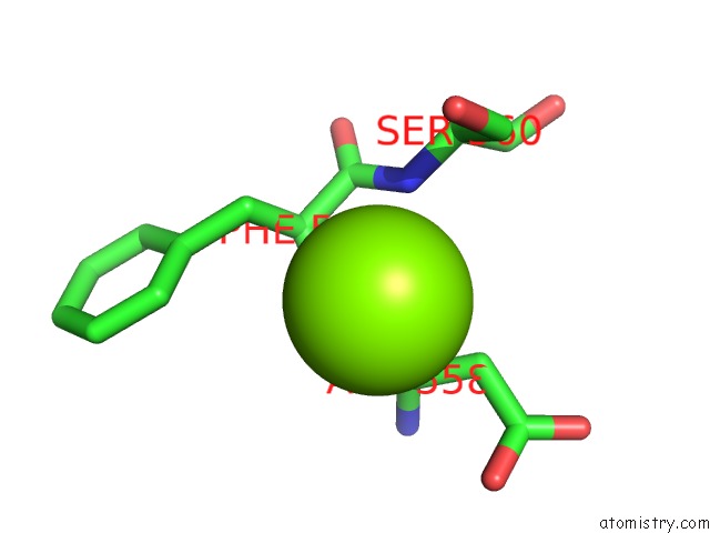
Magnesium binding site 3 out of 5 in 3shz
Go back to
Magnesium binding site 3 out
of 5 in the Crystal Structure of the PDE5A1 Catalytic Domain in Complex with Novel Inhibitors
Mono view
Stereo pair view
Mono view
Stereo pair view
A full contact list of Magnesium with other atoms in the Mg binding
site number 3 of Crystal Structure of the PDE5A1 Catalytic Domain in Complex with Novel Inhibitors within 5.0Å range:
|
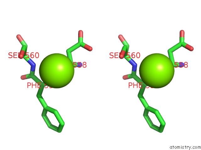
Magnesium binding site 4 out of 5 in 3shz
Go back to
Magnesium binding site 4 out
of 5 in the Crystal Structure of the PDE5A1 Catalytic Domain in Complex with Novel Inhibitors
Mono view
Stereo pair view
Mono view
Stereo pair view
A full contact list of Magnesium with other atoms in the Mg binding
site number 4 of Crystal Structure of the PDE5A1 Catalytic Domain in Complex with Novel Inhibitors within 5.0Å range:
|
Magnesium binding site 5 out of 5 in 3shz
Go back to
Magnesium binding site 5 out
of 5 in the Crystal Structure of the PDE5A1 Catalytic Domain in Complex with Novel Inhibitors
Mono view
Stereo pair view
Mono view
Stereo pair view
A full contact list of Magnesium with other atoms in the Mg binding
site number 5 of Crystal Structure of the PDE5A1 Catalytic Domain in Complex with Novel Inhibitors within 5.0Å range:
|
Reference:
Z.Xu,
Z.Liu,
T.Chen,
T.T.Chen,
Z.Wang,
G.Tian,
J.Shi,
X.Wang,
Y.Lu,
X.Yan,
G.Wang,
H.Jiang,
K.Chen,
S.Wang,
Y.Xu,
J.Shen,
W.Zhu.
Utilization of Halogen Bond in Lead Optimization: A Case Study of Rational Design of Potent Phosphodiesterase Type 5 (PDE5) Inhibitors. J.Med.Chem. V. 54 5607 2011.
ISSN: ISSN 0022-2623
PubMed: 21714539
DOI: 10.1021/JM200644R
Page generated: Thu Aug 15 10:59:27 2024
ISSN: ISSN 0022-2623
PubMed: 21714539
DOI: 10.1021/JM200644R
Last articles
K in 8Q0OK in 8Q0M
K in 8Q0J
K in 8PMK
K in 8Q0F
K in 8P4P
K in 8P4R
K in 8Q0A
K in 8PPM
K in 8PJL