Magnesium »
PDB 3siy-3srf »
3skl »
Magnesium in PDB 3skl: Crystal Structure of the 2'- Deoxyguanosine Riboswitch Bound to 2'- Deoxyguanosine, Iridium Hexammine Soak
Protein crystallography data
The structure of Crystal Structure of the 2'- Deoxyguanosine Riboswitch Bound to 2'- Deoxyguanosine, Iridium Hexammine Soak, PDB code: 3skl
was solved by
O.Pikovskaya,
A.Polonskaia,
D.J.Patel,
A.Serganov,
with X-Ray Crystallography technique. A brief refinement statistics is given in the table below:
| Resolution Low / High (Å) | 20.00 / 2.90 |
| Space group | C 1 2 1 |
| Cell size a, b, c (Å), α, β, γ (°) | 98.002, 34.955, 111.057, 90.00, 92.45, 90.00 |
| R / Rfree (%) | 21.9 / 26.4 |
Other elements in 3skl:
The structure of Crystal Structure of the 2'- Deoxyguanosine Riboswitch Bound to 2'- Deoxyguanosine, Iridium Hexammine Soak also contains other interesting chemical elements:
| Iridium | (Ir) | 10 atoms |
Magnesium Binding Sites:
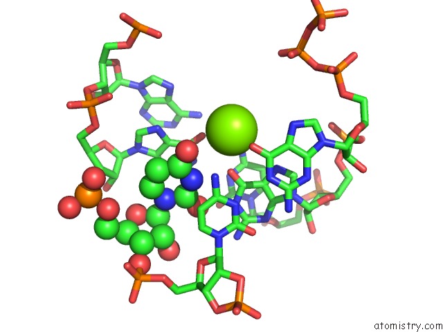
The binding sites of Magnesium atom in the Crystal Structure of the 2'- Deoxyguanosine Riboswitch Bound to 2'- Deoxyguanosine, Iridium Hexammine Soak
(pdb code 3skl). This binding sites where shown within
5.0 Angstroms radius around Magnesium atom.
In total 6 binding sites of Magnesium where determined in the Crystal Structure of the 2'- Deoxyguanosine Riboswitch Bound to 2'- Deoxyguanosine, Iridium Hexammine Soak, PDB code: 3skl:
Jump to Magnesium binding site number: 1; 2; 3; 4; 5; 6;
In total 6 binding sites of Magnesium where determined in the Crystal Structure of the 2'- Deoxyguanosine Riboswitch Bound to 2'- Deoxyguanosine, Iridium Hexammine Soak, PDB code: 3skl:
Jump to Magnesium binding site number: 1; 2; 3; 4; 5; 6;
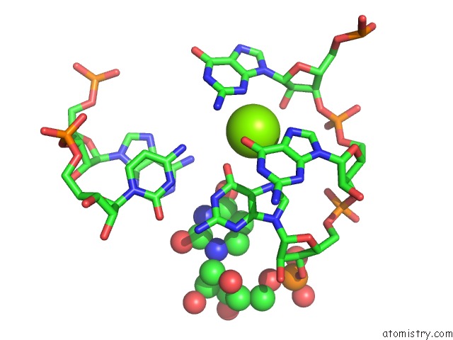
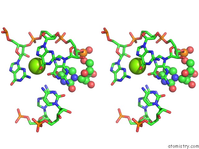
Magnesium binding site 1 out of 6 in 3skl
Go back to
Magnesium binding site 1 out
of 6 in the Crystal Structure of the 2'- Deoxyguanosine Riboswitch Bound to 2'- Deoxyguanosine, Iridium Hexammine Soak
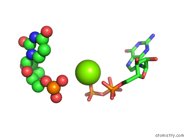
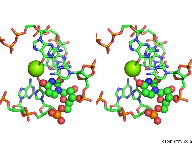
Mono view
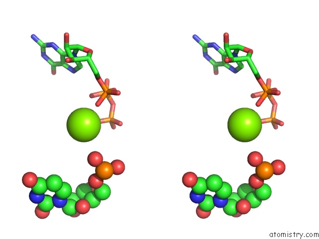
Stereo pair view
Mono view
Stereo pair view
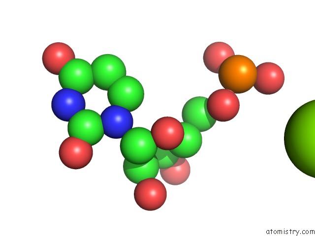
A full contact list of Magnesium with other atoms in the Mg binding
site number 1 of Crystal Structure of the 2'- Deoxyguanosine Riboswitch Bound to 2'- Deoxyguanosine, Iridium Hexammine Soak within 5.0Å range:
|
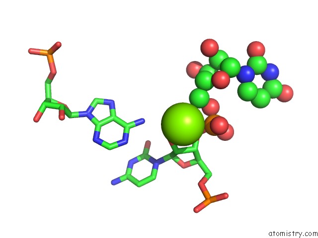
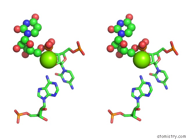
Magnesium binding site 2 out of 6 in 3skl
Go back to
Magnesium binding site 2 out
of 6 in the Crystal Structure of the 2'- Deoxyguanosine Riboswitch Bound to 2'- Deoxyguanosine, Iridium Hexammine Soak
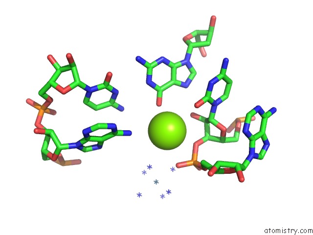
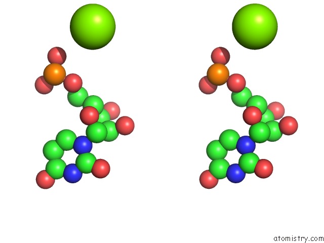
Mono view
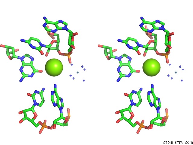
Stereo pair view
Mono view
Stereo pair view
A full contact list of Magnesium with other atoms in the Mg binding
site number 2 of Crystal Structure of the 2'- Deoxyguanosine Riboswitch Bound to 2'- Deoxyguanosine, Iridium Hexammine Soak within 5.0Å range:
|
Magnesium binding site 3 out of 6 in 3skl
Go back to
Magnesium binding site 3 out
of 6 in the Crystal Structure of the 2'- Deoxyguanosine Riboswitch Bound to 2'- Deoxyguanosine, Iridium Hexammine Soak
Mono view
Stereo pair view
Mono view
Stereo pair view
A full contact list of Magnesium with other atoms in the Mg binding
site number 3 of Crystal Structure of the 2'- Deoxyguanosine Riboswitch Bound to 2'- Deoxyguanosine, Iridium Hexammine Soak within 5.0Å range:
|
Magnesium binding site 4 out of 6 in 3skl
Go back to
Magnesium binding site 4 out
of 6 in the Crystal Structure of the 2'- Deoxyguanosine Riboswitch Bound to 2'- Deoxyguanosine, Iridium Hexammine Soak
Mono view
Stereo pair view
Mono view
Stereo pair view
A full contact list of Magnesium with other atoms in the Mg binding
site number 4 of Crystal Structure of the 2'- Deoxyguanosine Riboswitch Bound to 2'- Deoxyguanosine, Iridium Hexammine Soak within 5.0Å range:
|
Magnesium binding site 5 out of 6 in 3skl
Go back to
Magnesium binding site 5 out
of 6 in the Crystal Structure of the 2'- Deoxyguanosine Riboswitch Bound to 2'- Deoxyguanosine, Iridium Hexammine Soak
Mono view
Stereo pair view
Mono view
Stereo pair view
A full contact list of Magnesium with other atoms in the Mg binding
site number 5 of Crystal Structure of the 2'- Deoxyguanosine Riboswitch Bound to 2'- Deoxyguanosine, Iridium Hexammine Soak within 5.0Å range:
|
Magnesium binding site 6 out of 6 in 3skl
Go back to
Magnesium binding site 6 out
of 6 in the Crystal Structure of the 2'- Deoxyguanosine Riboswitch Bound to 2'- Deoxyguanosine, Iridium Hexammine Soak
Mono view
Stereo pair view
Mono view
Stereo pair view
A full contact list of Magnesium with other atoms in the Mg binding
site number 6 of Crystal Structure of the 2'- Deoxyguanosine Riboswitch Bound to 2'- Deoxyguanosine, Iridium Hexammine Soak within 5.0Å range:
|
Reference:
O.Pikovskaya,
A.Polonskaia,
D.J.Patel,
A.Serganov.
Structural Principles of Nucleoside Selectivity in A 2'-Deoxyguanosine Riboswitch. Nat.Chem.Biol. V. 7 748 2011.
ISSN: ISSN 1552-4450
PubMed: 21841796
DOI: 10.1038/NCHEMBIO.631
Page generated: Thu Aug 15 11:01:57 2024
ISSN: ISSN 1552-4450
PubMed: 21841796
DOI: 10.1038/NCHEMBIO.631
Last articles
Fe in 8BJ9Fe in 8BEW
Fe in 8BGW
Fe in 8BHX
Fe in 8BF6
Fe in 8BEE
Fe in 8BEL
Fe in 8BED
Fe in 8BCX
Fe in 8BEF