Magnesium »
PDB 3ss8-3t2c »
3st8 »
Magnesium in PDB 3st8: Crystal Structure of Glmu From Mycobacterium Tuberculosis in Complex with Coenzyme A, Glucosamine 1-Phosphate and Uridine-Diphosphate-N- Acetylglucosamine
Enzymatic activity of Crystal Structure of Glmu From Mycobacterium Tuberculosis in Complex with Coenzyme A, Glucosamine 1-Phosphate and Uridine-Diphosphate-N- Acetylglucosamine
All present enzymatic activity of Crystal Structure of Glmu From Mycobacterium Tuberculosis in Complex with Coenzyme A, Glucosamine 1-Phosphate and Uridine-Diphosphate-N- Acetylglucosamine:
2.3.1.157; 2.7.7.23;
2.3.1.157; 2.7.7.23;
Protein crystallography data
The structure of Crystal Structure of Glmu From Mycobacterium Tuberculosis in Complex with Coenzyme A, Glucosamine 1-Phosphate and Uridine-Diphosphate-N- Acetylglucosamine, PDB code: 3st8
was solved by
P.A.Jagtap,
B.Prakash,
with X-Ray Crystallography technique. A brief refinement statistics is given in the table below:
| Resolution Low / High (Å) | 19.71 / 1.98 |
| Space group | H 3 2 |
| Cell size a, b, c (Å), α, β, γ (°) | 110.312, 110.312, 360.537, 90.00, 90.00, 120.00 |
| R / Rfree (%) | 16.7 / 20.2 |
Magnesium Binding Sites:
The binding sites of Magnesium atom in the Crystal Structure of Glmu From Mycobacterium Tuberculosis in Complex with Coenzyme A, Glucosamine 1-Phosphate and Uridine-Diphosphate-N- Acetylglucosamine
(pdb code 3st8). This binding sites where shown within
5.0 Angstroms radius around Magnesium atom.
In total 4 binding sites of Magnesium where determined in the Crystal Structure of Glmu From Mycobacterium Tuberculosis in Complex with Coenzyme A, Glucosamine 1-Phosphate and Uridine-Diphosphate-N- Acetylglucosamine, PDB code: 3st8:
Jump to Magnesium binding site number: 1; 2; 3; 4;
In total 4 binding sites of Magnesium where determined in the Crystal Structure of Glmu From Mycobacterium Tuberculosis in Complex with Coenzyme A, Glucosamine 1-Phosphate and Uridine-Diphosphate-N- Acetylglucosamine, PDB code: 3st8:
Jump to Magnesium binding site number: 1; 2; 3; 4;
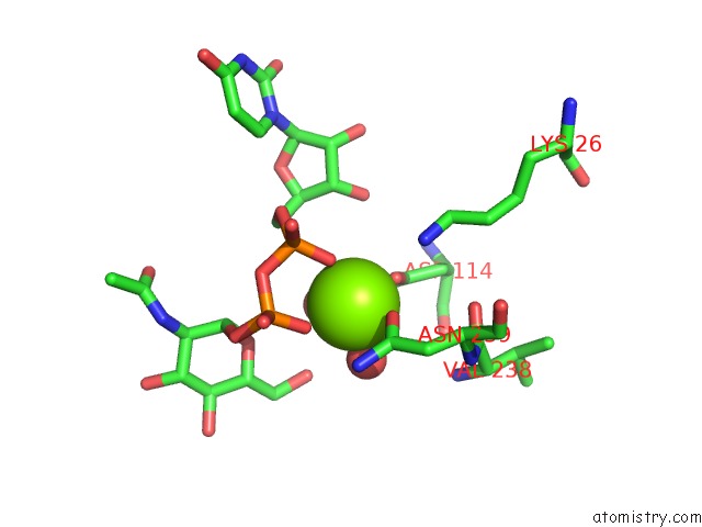
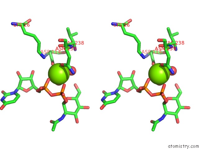
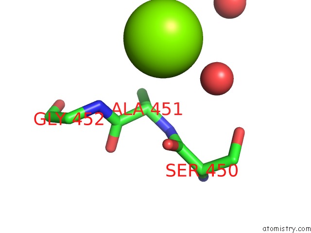
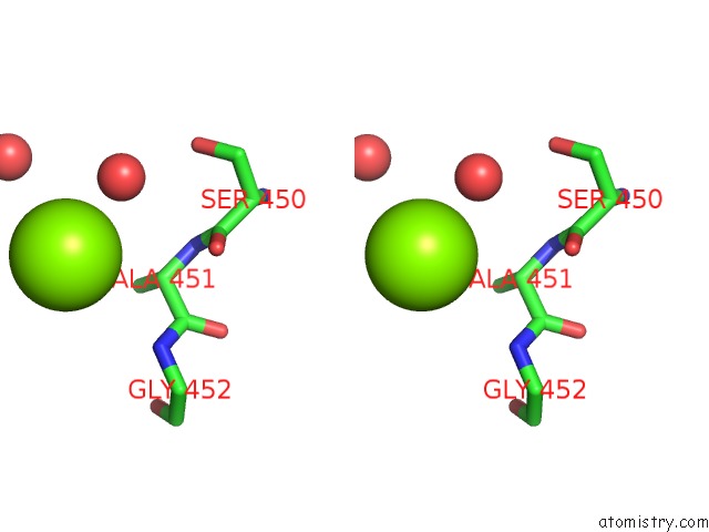
Magnesium binding site 1 out of 4 in 3st8
Go back to
Magnesium binding site 1 out
of 4 in the Crystal Structure of Glmu From Mycobacterium Tuberculosis in Complex with Coenzyme A, Glucosamine 1-Phosphate and Uridine-Diphosphate-N- Acetylglucosamine
Mono view
Stereo pair view
Mono view
Stereo pair view
A full contact list of Magnesium with other atoms in the Mg binding
site number 1 of Crystal Structure of Glmu From Mycobacterium Tuberculosis in Complex with Coenzyme A, Glucosamine 1-Phosphate and Uridine-Diphosphate-N- Acetylglucosamine within 5.0Å range:
|
Magnesium binding site 2 out of 4 in 3st8
Go back to
Magnesium binding site 2 out
of 4 in the Crystal Structure of Glmu From Mycobacterium Tuberculosis in Complex with Coenzyme A, Glucosamine 1-Phosphate and Uridine-Diphosphate-N- Acetylglucosamine
Mono view
Stereo pair view
Mono view
Stereo pair view
A full contact list of Magnesium with other atoms in the Mg binding
site number 2 of Crystal Structure of Glmu From Mycobacterium Tuberculosis in Complex with Coenzyme A, Glucosamine 1-Phosphate and Uridine-Diphosphate-N- Acetylglucosamine within 5.0Å range:
|
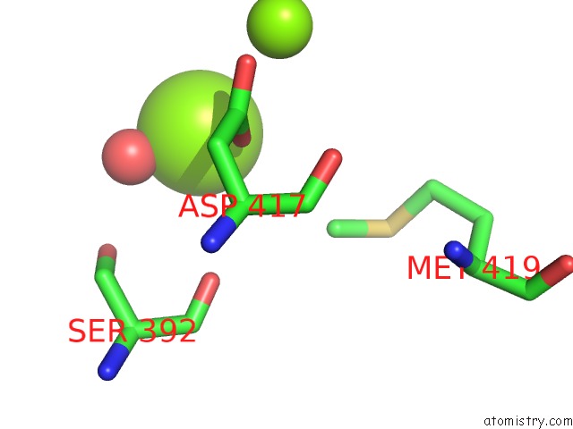
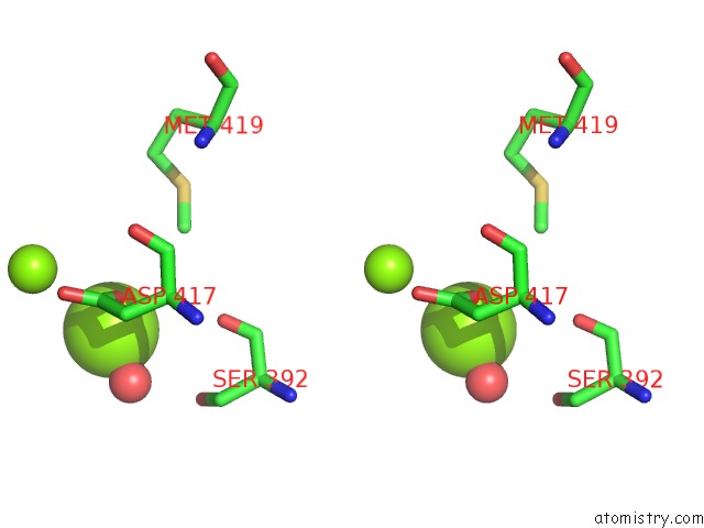
Magnesium binding site 3 out of 4 in 3st8
Go back to
Magnesium binding site 3 out
of 4 in the Crystal Structure of Glmu From Mycobacterium Tuberculosis in Complex with Coenzyme A, Glucosamine 1-Phosphate and Uridine-Diphosphate-N- Acetylglucosamine
Mono view
Stereo pair view
Mono view
Stereo pair view
A full contact list of Magnesium with other atoms in the Mg binding
site number 3 of Crystal Structure of Glmu From Mycobacterium Tuberculosis in Complex with Coenzyme A, Glucosamine 1-Phosphate and Uridine-Diphosphate-N- Acetylglucosamine within 5.0Å range:
|
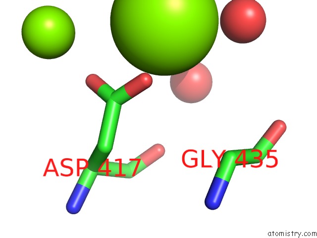
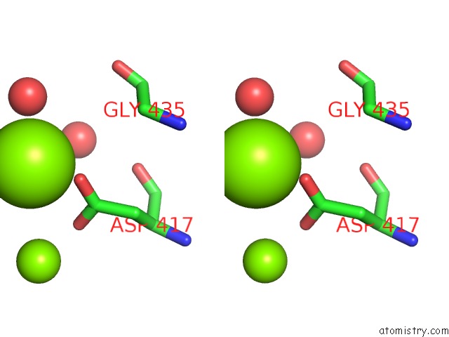
Magnesium binding site 4 out of 4 in 3st8
Go back to
Magnesium binding site 4 out
of 4 in the Crystal Structure of Glmu From Mycobacterium Tuberculosis in Complex with Coenzyme A, Glucosamine 1-Phosphate and Uridine-Diphosphate-N- Acetylglucosamine
Mono view
Stereo pair view
Mono view
Stereo pair view
A full contact list of Magnesium with other atoms in the Mg binding
site number 4 of Crystal Structure of Glmu From Mycobacterium Tuberculosis in Complex with Coenzyme A, Glucosamine 1-Phosphate and Uridine-Diphosphate-N- Acetylglucosamine within 5.0Å range:
|
Reference:
P.A.Jagtap,
B.Prakash.
Structure of Mycobacterium Tuberculosis Glmu in Complex with Acetyl Coa To Be Published.
Page generated: Mon Aug 11 03:14:49 2025
Last articles
Mg in 5O5JMg in 5NRG
Mg in 5O5W
Mg in 5O4V
Mg in 5O4C
Mg in 5O48
Mg in 5O44
Mg in 5O2S
Mg in 5O3W
Mg in 5O3V