Magnesium »
PDB 3ss8-3t2c »
3sz5 »
Magnesium in PDB 3sz5: Crystal Structure of Lhk-Exo in Complex with 5-Phosphorylated Oligothymidine (Dt)4
Protein crystallography data
The structure of Crystal Structure of Lhk-Exo in Complex with 5-Phosphorylated Oligothymidine (Dt)4, PDB code: 3sz5
was solved by
W.Yang,
W.Y.Chen,
H.Wang,
Q.Zhang,
W.Zhou,
M.Bartlam,
R.M.Watt,
Z.Rao,
with X-Ray Crystallography technique. A brief refinement statistics is given in the table below:
| Resolution Low / High (Å) | 50.00 / 2.80 |
| Space group | P 63 |
| Cell size a, b, c (Å), α, β, γ (°) | 108.697, 108.697, 48.219, 90.00, 90.00, 120.00 |
| R / Rfree (%) | 19.3 / 24.8 |
Magnesium Binding Sites:
The binding sites of Magnesium atom in the Crystal Structure of Lhk-Exo in Complex with 5-Phosphorylated Oligothymidine (Dt)4
(pdb code 3sz5). This binding sites where shown within
5.0 Angstroms radius around Magnesium atom.
In total 2 binding sites of Magnesium where determined in the Crystal Structure of Lhk-Exo in Complex with 5-Phosphorylated Oligothymidine (Dt)4, PDB code: 3sz5:
Jump to Magnesium binding site number: 1; 2;
In total 2 binding sites of Magnesium where determined in the Crystal Structure of Lhk-Exo in Complex with 5-Phosphorylated Oligothymidine (Dt)4, PDB code: 3sz5:
Jump to Magnesium binding site number: 1; 2;
Magnesium binding site 1 out of 2 in 3sz5
Go back to
Magnesium binding site 1 out
of 2 in the Crystal Structure of Lhk-Exo in Complex with 5-Phosphorylated Oligothymidine (Dt)4
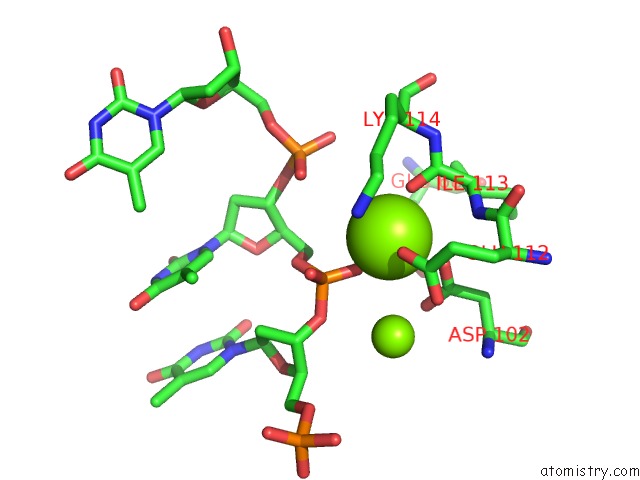
Mono view
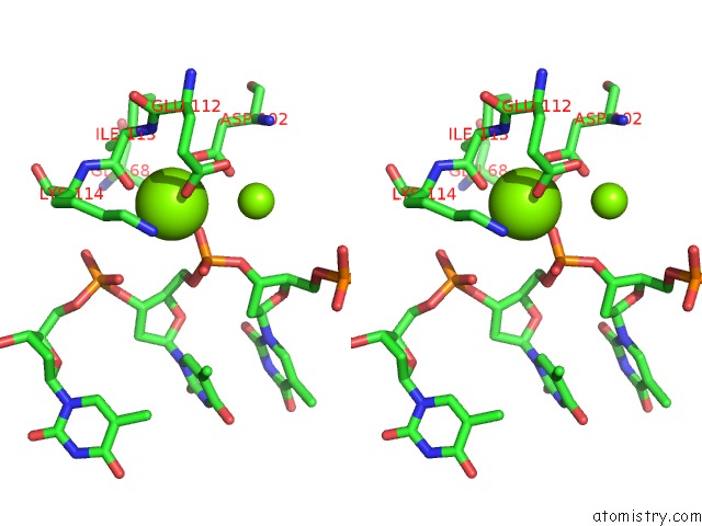
Stereo pair view
Mono view
Stereo pair view
A full contact list of Magnesium with other atoms in the Mg binding
site number 1 of Crystal Structure of Lhk-Exo in Complex with 5-Phosphorylated Oligothymidine (Dt)4 within 5.0Å range:
|
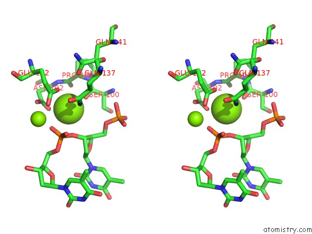
Magnesium binding site 2 out of 2 in 3sz5
Go back to
Magnesium binding site 2 out
of 2 in the Crystal Structure of Lhk-Exo in Complex with 5-Phosphorylated Oligothymidine (Dt)4

Mono view
Stereo pair view
Mono view
Stereo pair view
A full contact list of Magnesium with other atoms in the Mg binding
site number 2 of Crystal Structure of Lhk-Exo in Complex with 5-Phosphorylated Oligothymidine (Dt)4 within 5.0Å range:
|
Reference:
W.Yang,
W.Y.Chen,
H.Wang,
J.W.Ho,
J.D.Huang,
P.C.Woo,
S.K.Lau,
K.Y.Yuen,
Q.Zhang,
W.Zhou,
M.Bartlam,
R.M.Watt,
Z.Rao.
Structural and Functional Insight Into the Mechanism of An Alkaline Exonuclease From Laribacter Hongkongensis. Nucleic Acids Res. V. 39 9803 2011.
ISSN: ISSN 0305-1048
PubMed: 21893587
DOI: 10.1093/NAR/GKR660
Page generated: Mon Aug 11 03:16:50 2025
ISSN: ISSN 0305-1048
PubMed: 21893587
DOI: 10.1093/NAR/GKR660
Last articles
Mg in 5XN1Mg in 5XLZ
Mg in 5XLT
Mg in 5XM7
Mg in 5XM3
Mg in 5XLH
Mg in 5XKM
Mg in 5XLG
Mg in 5XKH
Mg in 5XLF