Magnesium »
PDB 3t2d-3tar »
3t9e »
Magnesium in PDB 3t9e: Crystal Structure of the Catalytic Domain of Human Diphosphoinositol Pentakisphosphate Kinase 2 (PPIP5K2) in Complex with Adp, 5-(Pp)-IP5 (5-IP7) and MGF3 (Transition State Mimic)
Enzymatic activity of Crystal Structure of the Catalytic Domain of Human Diphosphoinositol Pentakisphosphate Kinase 2 (PPIP5K2) in Complex with Adp, 5-(Pp)-IP5 (5-IP7) and MGF3 (Transition State Mimic)
All present enzymatic activity of Crystal Structure of the Catalytic Domain of Human Diphosphoinositol Pentakisphosphate Kinase 2 (PPIP5K2) in Complex with Adp, 5-(Pp)-IP5 (5-IP7) and MGF3 (Transition State Mimic):
2.7.4.24;
2.7.4.24;
Protein crystallography data
The structure of Crystal Structure of the Catalytic Domain of Human Diphosphoinositol Pentakisphosphate Kinase 2 (PPIP5K2) in Complex with Adp, 5-(Pp)-IP5 (5-IP7) and MGF3 (Transition State Mimic), PDB code: 3t9e
was solved by
H.Wang,
J.Falck,
T.M.T.Hall,
S.B.Shears,
with X-Ray Crystallography technique. A brief refinement statistics is given in the table below:
| Resolution Low / High (Å) | 37.50 / 1.90 |
| Space group | P 21 21 21 |
| Cell size a, b, c (Å), α, β, γ (°) | 89.070, 109.987, 41.338, 90.00, 90.00, 90.00 |
| R / Rfree (%) | 17.3 / 21.3 |
Other elements in 3t9e:
The structure of Crystal Structure of the Catalytic Domain of Human Diphosphoinositol Pentakisphosphate Kinase 2 (PPIP5K2) in Complex with Adp, 5-(Pp)-IP5 (5-IP7) and MGF3 (Transition State Mimic) also contains other interesting chemical elements:
| Fluorine | (F) | 3 atoms |
Magnesium Binding Sites:
The binding sites of Magnesium atom in the Crystal Structure of the Catalytic Domain of Human Diphosphoinositol Pentakisphosphate Kinase 2 (PPIP5K2) in Complex with Adp, 5-(Pp)-IP5 (5-IP7) and MGF3 (Transition State Mimic)
(pdb code 3t9e). This binding sites where shown within
5.0 Angstroms radius around Magnesium atom.
In total 6 binding sites of Magnesium where determined in the Crystal Structure of the Catalytic Domain of Human Diphosphoinositol Pentakisphosphate Kinase 2 (PPIP5K2) in Complex with Adp, 5-(Pp)-IP5 (5-IP7) and MGF3 (Transition State Mimic), PDB code: 3t9e:
Jump to Magnesium binding site number: 1; 2; 3; 4; 5; 6;
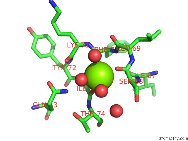
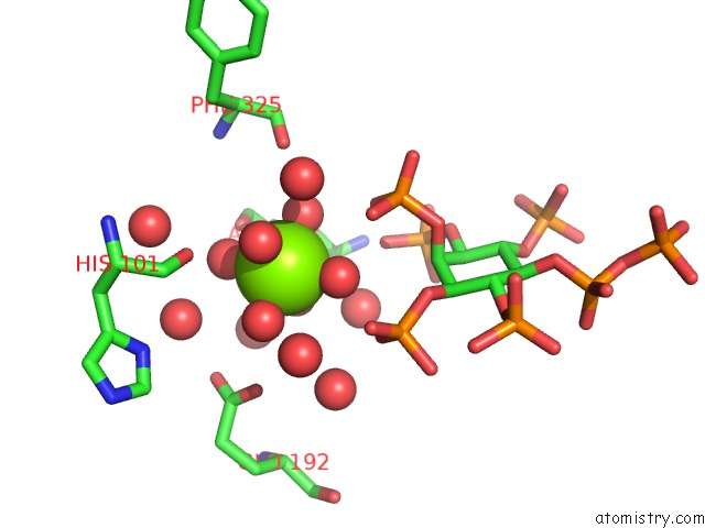
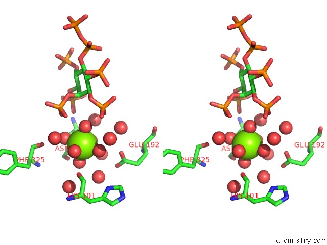
Magnesium binding site 1 out of 6 in 3t9e
Go back to
Magnesium binding site 1 out
of 6 in the Crystal Structure of the Catalytic Domain of Human Diphosphoinositol Pentakisphosphate Kinase 2 (PPIP5K2) in Complex with Adp, 5-(Pp)-IP5 (5-IP7) and MGF3 (Transition State Mimic)
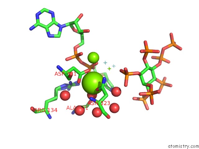
Mono view
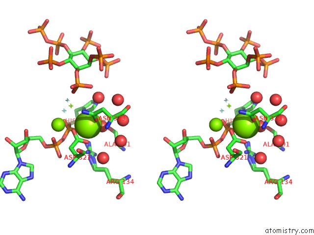
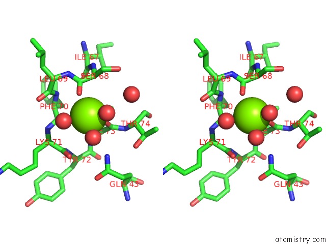
Stereo pair view
Mono view
Stereo pair view
A full contact list of Magnesium with other atoms in the Mg binding
site number 1 of Crystal Structure of the Catalytic Domain of Human Diphosphoinositol Pentakisphosphate Kinase 2 (PPIP5K2) in Complex with Adp, 5-(Pp)-IP5 (5-IP7) and MGF3 (Transition State Mimic) within 5.0Å range:
|
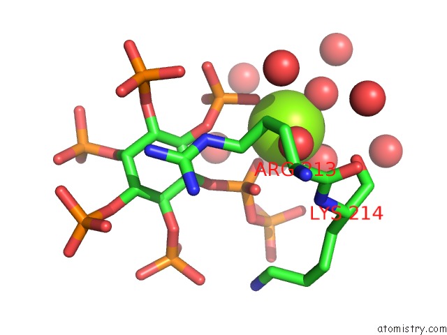
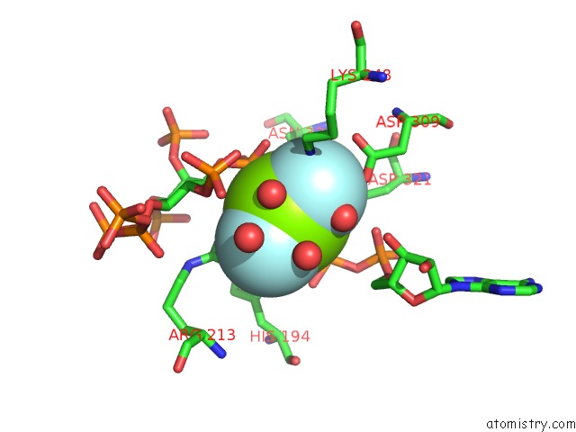
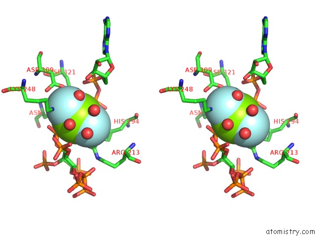
Magnesium binding site 2 out of 6 in 3t9e
Go back to
Magnesium binding site 2 out
of 6 in the Crystal Structure of the Catalytic Domain of Human Diphosphoinositol Pentakisphosphate Kinase 2 (PPIP5K2) in Complex with Adp, 5-(Pp)-IP5 (5-IP7) and MGF3 (Transition State Mimic)
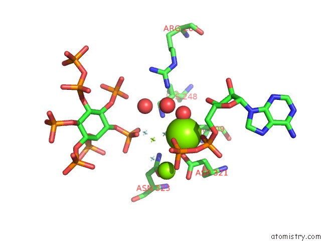
Mono view
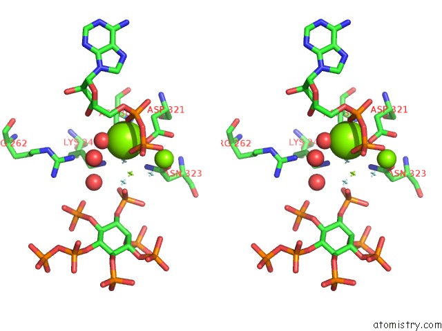
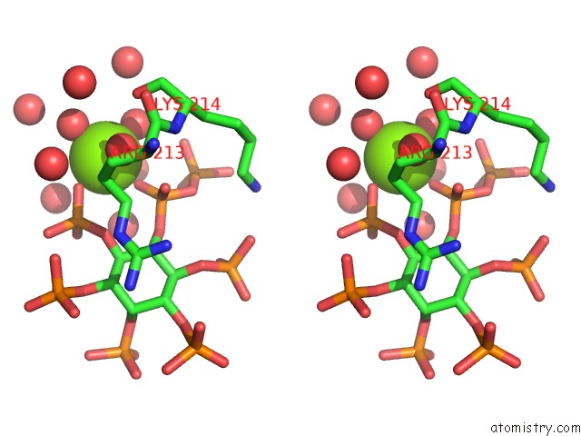
Stereo pair view
Mono view
Stereo pair view
A full contact list of Magnesium with other atoms in the Mg binding
site number 2 of Crystal Structure of the Catalytic Domain of Human Diphosphoinositol Pentakisphosphate Kinase 2 (PPIP5K2) in Complex with Adp, 5-(Pp)-IP5 (5-IP7) and MGF3 (Transition State Mimic) within 5.0Å range:
|
Magnesium binding site 3 out of 6 in 3t9e
Go back to
Magnesium binding site 3 out
of 6 in the Crystal Structure of the Catalytic Domain of Human Diphosphoinositol Pentakisphosphate Kinase 2 (PPIP5K2) in Complex with Adp, 5-(Pp)-IP5 (5-IP7) and MGF3 (Transition State Mimic)
Mono view
Stereo pair view
Mono view
Stereo pair view
A full contact list of Magnesium with other atoms in the Mg binding
site number 3 of Crystal Structure of the Catalytic Domain of Human Diphosphoinositol Pentakisphosphate Kinase 2 (PPIP5K2) in Complex with Adp, 5-(Pp)-IP5 (5-IP7) and MGF3 (Transition State Mimic) within 5.0Å range:
|
Magnesium binding site 4 out of 6 in 3t9e
Go back to
Magnesium binding site 4 out
of 6 in the Crystal Structure of the Catalytic Domain of Human Diphosphoinositol Pentakisphosphate Kinase 2 (PPIP5K2) in Complex with Adp, 5-(Pp)-IP5 (5-IP7) and MGF3 (Transition State Mimic)
Mono view
Stereo pair view
Mono view
Stereo pair view
A full contact list of Magnesium with other atoms in the Mg binding
site number 4 of Crystal Structure of the Catalytic Domain of Human Diphosphoinositol Pentakisphosphate Kinase 2 (PPIP5K2) in Complex with Adp, 5-(Pp)-IP5 (5-IP7) and MGF3 (Transition State Mimic) within 5.0Å range:
|
Magnesium binding site 5 out of 6 in 3t9e
Go back to
Magnesium binding site 5 out
of 6 in the Crystal Structure of the Catalytic Domain of Human Diphosphoinositol Pentakisphosphate Kinase 2 (PPIP5K2) in Complex with Adp, 5-(Pp)-IP5 (5-IP7) and MGF3 (Transition State Mimic)
Mono view
Stereo pair view
Mono view
Stereo pair view
A full contact list of Magnesium with other atoms in the Mg binding
site number 5 of Crystal Structure of the Catalytic Domain of Human Diphosphoinositol Pentakisphosphate Kinase 2 (PPIP5K2) in Complex with Adp, 5-(Pp)-IP5 (5-IP7) and MGF3 (Transition State Mimic) within 5.0Å range:
|
Magnesium binding site 6 out of 6 in 3t9e
Go back to
Magnesium binding site 6 out
of 6 in the Crystal Structure of the Catalytic Domain of Human Diphosphoinositol Pentakisphosphate Kinase 2 (PPIP5K2) in Complex with Adp, 5-(Pp)-IP5 (5-IP7) and MGF3 (Transition State Mimic)
Mono view
Stereo pair view
Mono view
Stereo pair view
A full contact list of Magnesium with other atoms in the Mg binding
site number 6 of Crystal Structure of the Catalytic Domain of Human Diphosphoinositol Pentakisphosphate Kinase 2 (PPIP5K2) in Complex with Adp, 5-(Pp)-IP5 (5-IP7) and MGF3 (Transition State Mimic) within 5.0Å range:
|
Reference:
H.Wang,
J.R.Falck,
T.M.Hall,
S.B.Shears.
Structural Basis For An Inositol Pyrophosphate Kinase Surmounting Phosphate Crowding. Nat.Chem.Biol. V. 8 111 2011.
ISSN: ISSN 1552-4450
PubMed: 22119861
DOI: 10.1038/NCHEMBIO.733
Page generated: Thu Aug 15 11:59:51 2024
ISSN: ISSN 1552-4450
PubMed: 22119861
DOI: 10.1038/NCHEMBIO.733
Last articles
Mg in 3T2CMg in 3T2B
Mg in 3T1R
Mg in 3T1O
Mg in 3T0D
Mg in 3T1Q
Mg in 3T12
Mg in 3T1K
Mg in 3T10
Mg in 3T0Z