Magnesium »
PDB 3tnq-3twp »
3to3 »
Magnesium in PDB 3to3: Crystal Structure of Petrobactin Biosynthesis Protein Asbb From Bacillus Anthracis Str. Sterne
Protein crystallography data
The structure of Crystal Structure of Petrobactin Biosynthesis Protein Asbb From Bacillus Anthracis Str. Sterne, PDB code: 3to3
was solved by
Y.Kim,
W.Eschenfeldt,
L.Stols,
A.Joachimiak,
Midwest Center Forstructural Genomics (Mcsg),
with X-Ray Crystallography technique. A brief refinement statistics is given in the table below:
| Resolution Low / High (Å) | 49.63 / 2.38 |
| Space group | P 21 21 21 |
| Cell size a, b, c (Å), α, β, γ (°) | 64.259, 155.923, 156.244, 90.00, 90.00, 90.00 |
| R / Rfree (%) | 17.2 / 22.7 |
Other elements in 3to3:
The structure of Crystal Structure of Petrobactin Biosynthesis Protein Asbb From Bacillus Anthracis Str. Sterne also contains other interesting chemical elements:
| Chlorine | (Cl) | 2 atoms |
Magnesium Binding Sites:
The binding sites of Magnesium atom in the Crystal Structure of Petrobactin Biosynthesis Protein Asbb From Bacillus Anthracis Str. Sterne
(pdb code 3to3). This binding sites where shown within
5.0 Angstroms radius around Magnesium atom.
In total only one binding site of Magnesium was determined in the Crystal Structure of Petrobactin Biosynthesis Protein Asbb From Bacillus Anthracis Str. Sterne, PDB code: 3to3:
In total only one binding site of Magnesium was determined in the Crystal Structure of Petrobactin Biosynthesis Protein Asbb From Bacillus Anthracis Str. Sterne, PDB code: 3to3:
Magnesium binding site 1 out of 1 in 3to3
Go back to
Magnesium binding site 1 out
of 1 in the Crystal Structure of Petrobactin Biosynthesis Protein Asbb From Bacillus Anthracis Str. Sterne
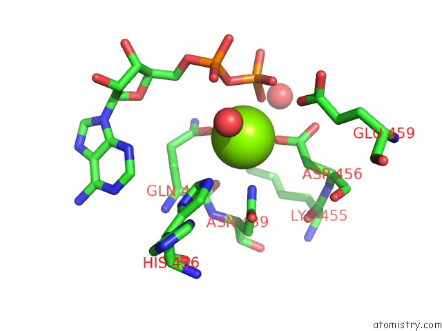
Mono view
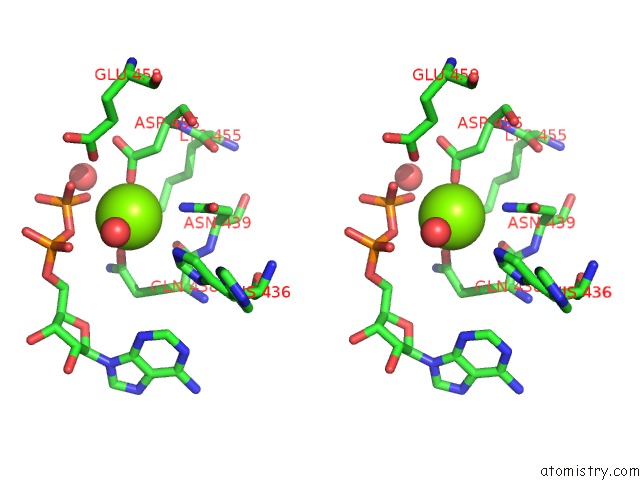
Stereo pair view
Mono view
Stereo pair view
A full contact list of Magnesium with other atoms in the Mg binding
site number 1 of Crystal Structure of Petrobactin Biosynthesis Protein Asbb From Bacillus Anthracis Str. Sterne within 5.0Å range:
|
Reference:
T.D.Nusca,
Y.Kim,
N.Maltseva,
J.Y.Lee,
W.Eschenfeldt,
L.Stols,
M.M.Schofield,
J.B.Scaglione,
S.D.Dixon,
D.Oves-Costales,
G.L.Challis,
P.C.Hanna,
B.F.Pfleger,
A.Joachimiak,
D.H.Sherman.
Functional and Structural Analysis of the Siderophore Synthetase Asbb Through Reconstitution of the Petrobactin Biosynthetic Pathway From Bacillus Anthracis. J.Biol.Chem. V. 287 16058 2012.
ISSN: ISSN 0021-9258
PubMed: 22408253
DOI: 10.1074/JBC.M112.359349
Page generated: Mon Aug 11 03:58:17 2025
ISSN: ISSN 0021-9258
PubMed: 22408253
DOI: 10.1074/JBC.M112.359349
Last articles
Mg in 4DTHMg in 4DTF
Mg in 4DSU
Mg in 4DST
Mg in 4DSO
Mg in 4DSN
Mg in 4DSC
Mg in 4DS6
Mg in 4DRX
Mg in 4DR4