Magnesium »
PDB 3tx9-3u8u »
3u67 »
Magnesium in PDB 3u67: Crystal Structure of the N-Terminal Domain of HSP90 From Leishmania Major(LMJF33.0312)in Complex with Adp
Protein crystallography data
The structure of Crystal Structure of the N-Terminal Domain of HSP90 From Leishmania Major(LMJF33.0312)in Complex with Adp, PDB code: 3u67
was solved by
J.C.Pizarro,
A.K.Wernimont,
A.Hutchinson,
F.Mackenzie,
A.Fairlamb,
C.H.Arrowsmith,
C.Bountra,
J.Weigelt,
A.M.Edwards,
M.A.J.Ferguson,
R.Hui,
T.Hills,
Structural Genomics Consortium (Sgc),
with X-Ray Crystallography technique. A brief refinement statistics is given in the table below:
| Resolution Low / High (Å) | 32.94 / 1.77 |
| Space group | P 21 21 2 |
| Cell size a, b, c (Å), α, β, γ (°) | 121.865, 42.991, 51.253, 90.00, 90.00, 90.00 |
| R / Rfree (%) | 20.2 / 24 |
Magnesium Binding Sites:
The binding sites of Magnesium atom in the Crystal Structure of the N-Terminal Domain of HSP90 From Leishmania Major(LMJF33.0312)in Complex with Adp
(pdb code 3u67). This binding sites where shown within
5.0 Angstroms radius around Magnesium atom.
In total only one binding site of Magnesium was determined in the Crystal Structure of the N-Terminal Domain of HSP90 From Leishmania Major(LMJF33.0312)in Complex with Adp, PDB code: 3u67:
In total only one binding site of Magnesium was determined in the Crystal Structure of the N-Terminal Domain of HSP90 From Leishmania Major(LMJF33.0312)in Complex with Adp, PDB code: 3u67:
Magnesium binding site 1 out of 1 in 3u67
Go back to
Magnesium binding site 1 out
of 1 in the Crystal Structure of the N-Terminal Domain of HSP90 From Leishmania Major(LMJF33.0312)in Complex with Adp
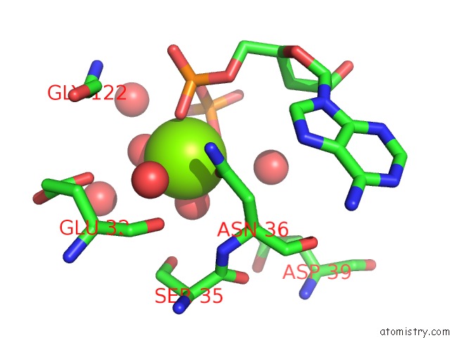
Mono view
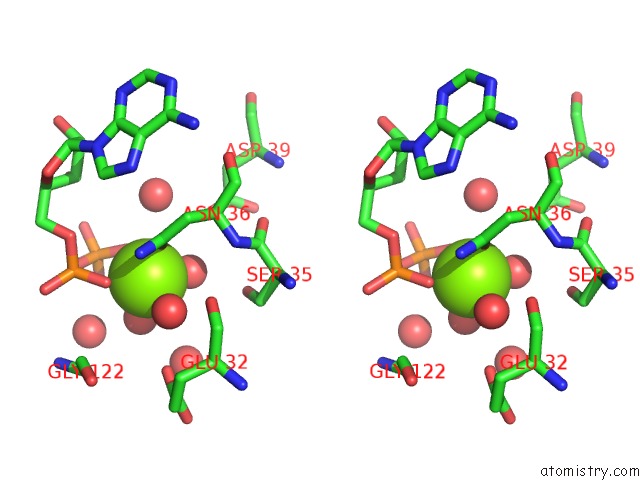
Stereo pair view
Mono view
Stereo pair view
A full contact list of Magnesium with other atoms in the Mg binding
site number 1 of Crystal Structure of the N-Terminal Domain of HSP90 From Leishmania Major(LMJF33.0312)in Complex with Adp within 5.0Å range:
|
Reference:
J.C.Pizarro,
T.Hills,
G.Senisterra,
A.K.Wernimont,
C.Mackenzie,
N.R.Norcross,
M.A.Ferguson,
P.G.Wyatt,
I.H.Gilbert,
R.Hui.
Exploring the Trypanosoma Brucei HSP83 Potential As A Target For Structure Guided Drug Design. Plos Negl Trop Dis V. 7 E2492 2013.
ISSN: ESSN 1935-2735
PubMed: 24147171
DOI: 10.1371/JOURNAL.PNTD.0002492
Page generated: Thu Aug 15 12:22:53 2024
ISSN: ESSN 1935-2735
PubMed: 24147171
DOI: 10.1371/JOURNAL.PNTD.0002492
Last articles
Mg in 3IJQMg in 3IJL
Mg in 3IJI
Mg in 3IJH
Mg in 3IIV
Mg in 3IIU
Mg in 3IIL
Mg in 3IIS
Mg in 3IHK
Mg in 3II9