Magnesium »
PDB 3umy-3v4i »
3uxl »
Magnesium in PDB 3uxl: P. Putida Mandelate Racemase Co-Crystallized with the Intermediate Analogue Cupferron
Enzymatic activity of P. Putida Mandelate Racemase Co-Crystallized with the Intermediate Analogue Cupferron
All present enzymatic activity of P. Putida Mandelate Racemase Co-Crystallized with the Intermediate Analogue Cupferron:
5.1.2.2;
5.1.2.2;
Protein crystallography data
The structure of P. Putida Mandelate Racemase Co-Crystallized with the Intermediate Analogue Cupferron, PDB code: 3uxl
was solved by
A.D.Lietzan,
E.Pellmann,
M.St Maurice,
with X-Ray Crystallography technique. A brief refinement statistics is given in the table below:
| Resolution Low / High (Å) | 50.00 / 2.20 |
| Space group | I 4 |
| Cell size a, b, c (Å), α, β, γ (°) | 148.256, 148.256, 175.388, 90.00, 90.00, 90.00 |
| R / Rfree (%) | 18.2 / 22.7 |
Magnesium Binding Sites:
The binding sites of Magnesium atom in the P. Putida Mandelate Racemase Co-Crystallized with the Intermediate Analogue Cupferron
(pdb code 3uxl). This binding sites where shown within
5.0 Angstroms radius around Magnesium atom.
In total 4 binding sites of Magnesium where determined in the P. Putida Mandelate Racemase Co-Crystallized with the Intermediate Analogue Cupferron, PDB code: 3uxl:
Jump to Magnesium binding site number: 1; 2; 3; 4;
In total 4 binding sites of Magnesium where determined in the P. Putida Mandelate Racemase Co-Crystallized with the Intermediate Analogue Cupferron, PDB code: 3uxl:
Jump to Magnesium binding site number: 1; 2; 3; 4;
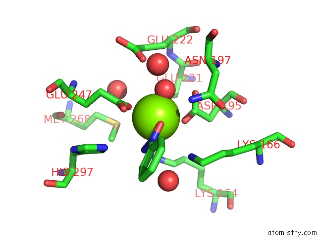
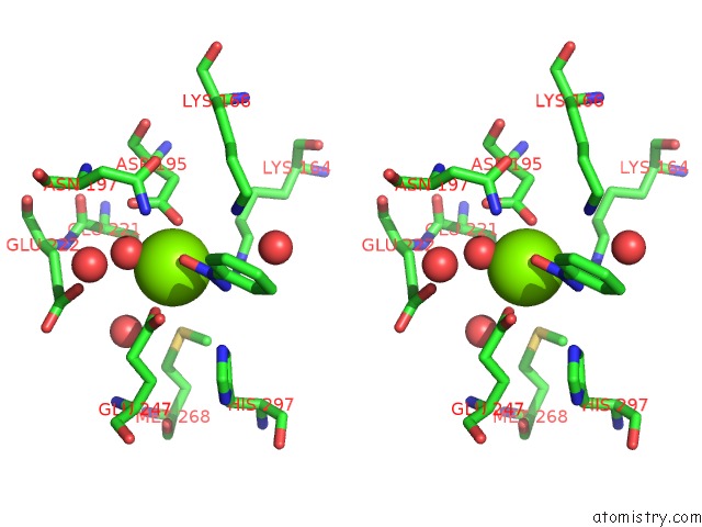
Magnesium binding site 1 out of 4 in 3uxl
Go back to
Magnesium binding site 1 out
of 4 in the P. Putida Mandelate Racemase Co-Crystallized with the Intermediate Analogue Cupferron
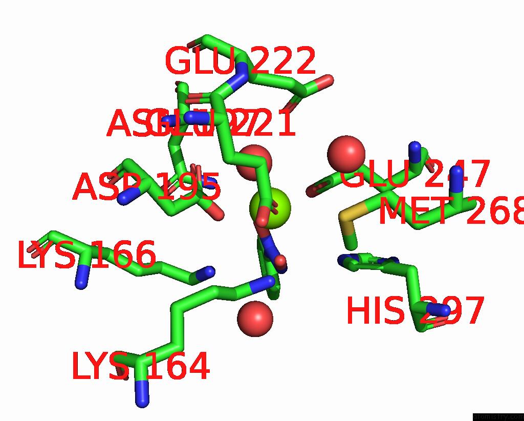
Mono view
Stereo pair view
Mono view
Stereo pair view
A full contact list of Magnesium with other atoms in the Mg binding
site number 1 of P. Putida Mandelate Racemase Co-Crystallized with the Intermediate Analogue Cupferron within 5.0Å range:
|
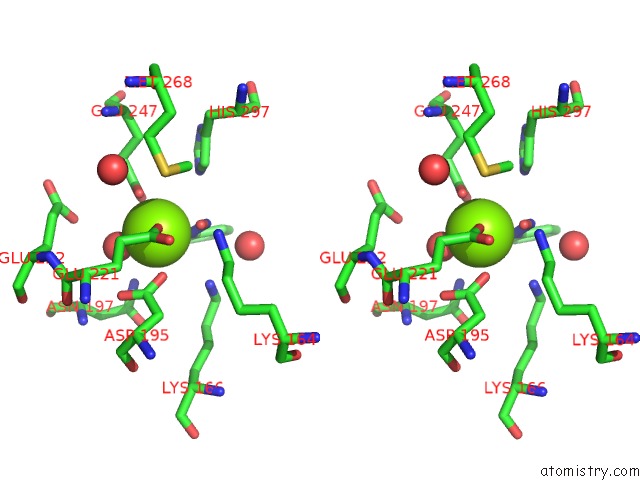
Magnesium binding site 2 out of 4 in 3uxl
Go back to
Magnesium binding site 2 out
of 4 in the P. Putida Mandelate Racemase Co-Crystallized with the Intermediate Analogue Cupferron
Mono view
Stereo pair view
Mono view
Stereo pair view
A full contact list of Magnesium with other atoms in the Mg binding
site number 2 of P. Putida Mandelate Racemase Co-Crystallized with the Intermediate Analogue Cupferron within 5.0Å range:
|
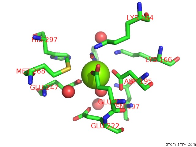
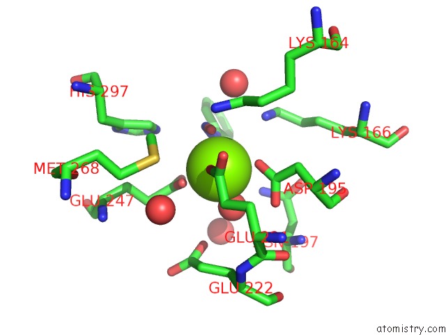
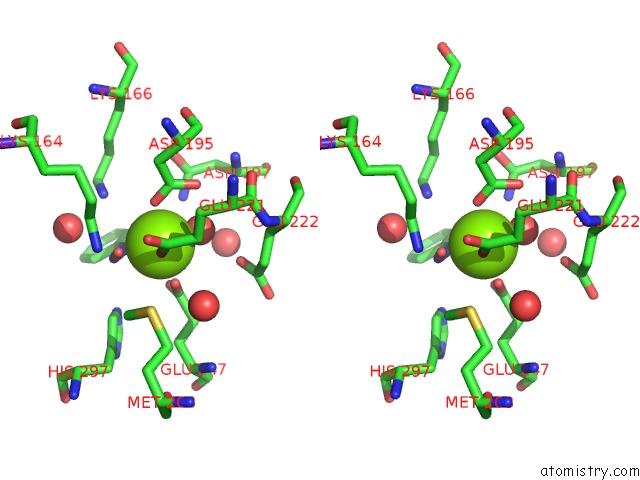
Magnesium binding site 3 out of 4 in 3uxl
Go back to
Magnesium binding site 3 out
of 4 in the P. Putida Mandelate Racemase Co-Crystallized with the Intermediate Analogue Cupferron
Mono view
Stereo pair view
Mono view
Stereo pair view
A full contact list of Magnesium with other atoms in the Mg binding
site number 3 of P. Putida Mandelate Racemase Co-Crystallized with the Intermediate Analogue Cupferron within 5.0Å range:
|
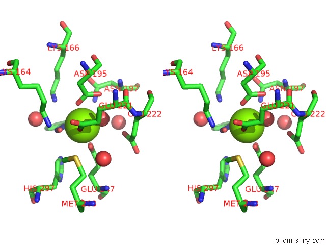
Magnesium binding site 4 out of 4 in 3uxl
Go back to
Magnesium binding site 4 out
of 4 in the P. Putida Mandelate Racemase Co-Crystallized with the Intermediate Analogue Cupferron
Mono view
Stereo pair view
Mono view
Stereo pair view
A full contact list of Magnesium with other atoms in the Mg binding
site number 4 of P. Putida Mandelate Racemase Co-Crystallized with the Intermediate Analogue Cupferron within 5.0Å range:
|
Reference:
A.D.Lietzan,
M.Nagar,
E.A.Pellmann,
J.R.Bourque,
S.L.Bearne,
M.St Maurice.
Structure of Mandelate Racemase with Bound Intermediate Analogues Benzohydroxamate and Cupferron. Biochemistry V. 51 1160 2012.
ISSN: ISSN 0006-2960
PubMed: 22264153
DOI: 10.1021/BI2018514
Page generated: Mon Aug 11 04:20:35 2025
ISSN: ISSN 0006-2960
PubMed: 22264153
DOI: 10.1021/BI2018514
Last articles
Mg in 5J8PMg in 5J8N
Mg in 5J78
Mg in 5J85
Mg in 5J5Q
Mg in 5J5P
Mg in 5J7R
Mg in 5J6O
Mg in 5J3Z
Mg in 5J33