Magnesium »
PDB 3vdc-3vth »
3vdi »
Magnesium in PDB 3vdi: Structure of the Fmo Protein From Pelodictyon Phaeum
Protein crystallography data
The structure of Structure of the Fmo Protein From Pelodictyon Phaeum, PDB code: 3vdi
was solved by
D.E.Tronrud,
C.R.Larson,
C.O.Seng,
L.Lauman,
H.J.Matthies,
J.Wen,
R.E.Blankenship,
J.P.Allen,
with X-Ray Crystallography technique. A brief refinement statistics is given in the table below:
| Resolution Low / High (Å) | 40.00 / 1.99 |
| Space group | P 63 |
| Cell size a, b, c (Å), α, β, γ (°) | 83.967, 83.967, 115.724, 90.00, 90.00, 120.00 |
| R / Rfree (%) | 16.2 / 19 |
Magnesium Binding Sites:
The binding sites of Magnesium atom in the Structure of the Fmo Protein From Pelodictyon Phaeum
(pdb code 3vdi). This binding sites where shown within
5.0 Angstroms radius around Magnesium atom.
In total 7 binding sites of Magnesium where determined in the Structure of the Fmo Protein From Pelodictyon Phaeum, PDB code: 3vdi:
Jump to Magnesium binding site number: 1; 2; 3; 4; 5; 6; 7;
In total 7 binding sites of Magnesium where determined in the Structure of the Fmo Protein From Pelodictyon Phaeum, PDB code: 3vdi:
Jump to Magnesium binding site number: 1; 2; 3; 4; 5; 6; 7;
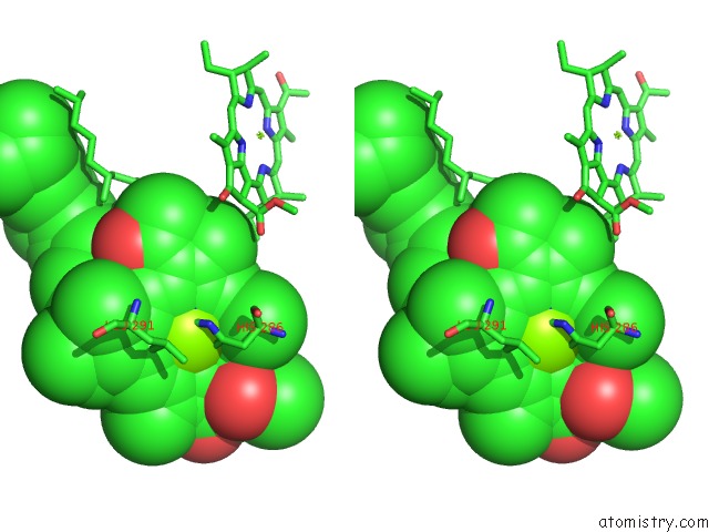
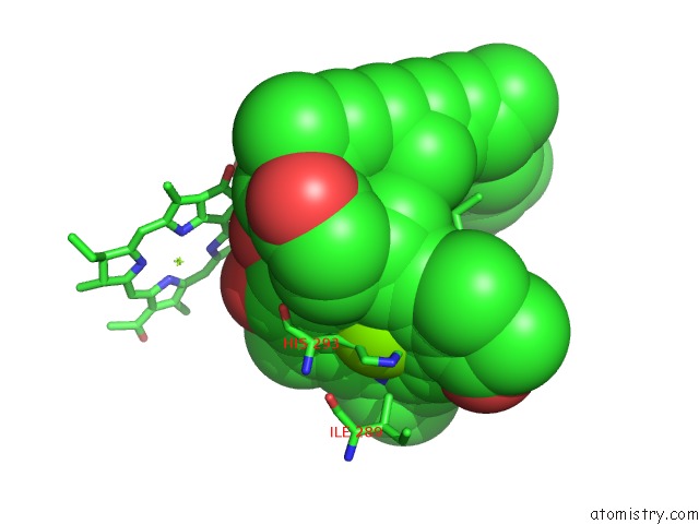
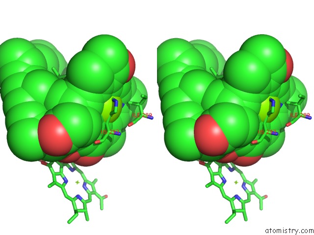
Magnesium binding site 1 out of 7 in 3vdi
Go back to
Magnesium binding site 1 out
of 7 in the Structure of the Fmo Protein From Pelodictyon Phaeum

Mono view
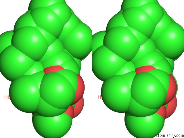
Stereo pair view
Mono view
Stereo pair view
A full contact list of Magnesium with other atoms in the Mg binding
site number 1 of Structure of the Fmo Protein From Pelodictyon Phaeum within 5.0Å range:
|
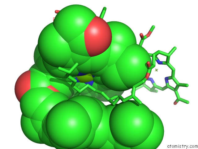
Magnesium binding site 2 out of 7 in 3vdi
Go back to
Magnesium binding site 2 out
of 7 in the Structure of the Fmo Protein From Pelodictyon Phaeum
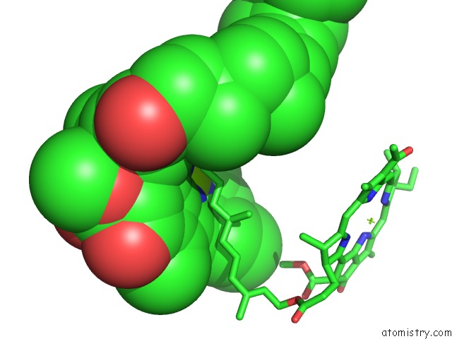
Mono view

Stereo pair view
Mono view
Stereo pair view
A full contact list of Magnesium with other atoms in the Mg binding
site number 2 of Structure of the Fmo Protein From Pelodictyon Phaeum within 5.0Å range:
|
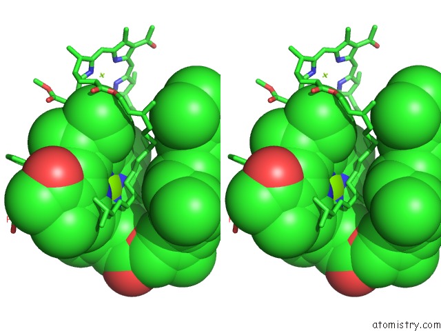
Magnesium binding site 3 out of 7 in 3vdi
Go back to
Magnesium binding site 3 out
of 7 in the Structure of the Fmo Protein From Pelodictyon Phaeum

Mono view
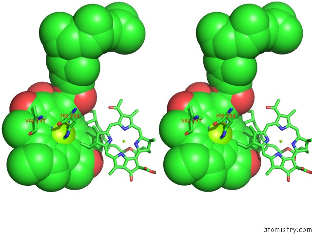
Stereo pair view
Mono view
Stereo pair view
A full contact list of Magnesium with other atoms in the Mg binding
site number 3 of Structure of the Fmo Protein From Pelodictyon Phaeum within 5.0Å range:
|
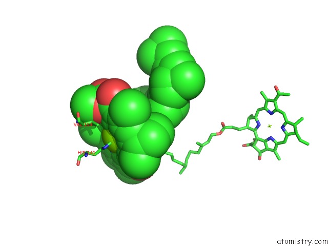
Magnesium binding site 4 out of 7 in 3vdi
Go back to
Magnesium binding site 4 out
of 7 in the Structure of the Fmo Protein From Pelodictyon Phaeum
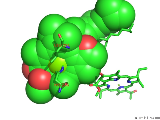
Mono view
Stereo pair view
Mono view
Stereo pair view
A full contact list of Magnesium with other atoms in the Mg binding
site number 4 of Structure of the Fmo Protein From Pelodictyon Phaeum within 5.0Å range:
|
Magnesium binding site 5 out of 7 in 3vdi
Go back to
Magnesium binding site 5 out
of 7 in the Structure of the Fmo Protein From Pelodictyon Phaeum
Mono view
Stereo pair view
Mono view
Stereo pair view
A full contact list of Magnesium with other atoms in the Mg binding
site number 5 of Structure of the Fmo Protein From Pelodictyon Phaeum within 5.0Å range:
|
Magnesium binding site 6 out of 7 in 3vdi
Go back to
Magnesium binding site 6 out
of 7 in the Structure of the Fmo Protein From Pelodictyon Phaeum
Mono view

Stereo pair view
Mono view
Stereo pair view
A full contact list of Magnesium with other atoms in the Mg binding
site number 6 of Structure of the Fmo Protein From Pelodictyon Phaeum within 5.0Å range:
|
Magnesium binding site 7 out of 7 in 3vdi
Go back to
Magnesium binding site 7 out
of 7 in the Structure of the Fmo Protein From Pelodictyon Phaeum
Mono view
Stereo pair view
Mono view
Stereo pair view
A full contact list of Magnesium with other atoms in the Mg binding
site number 7 of Structure of the Fmo Protein From Pelodictyon Phaeum within 5.0Å range:
|
Reference:
D.E.Tronrud,
J.P.Allen.
Reinterpretation of the Electron Density at the Site of the Eighth Bacteriochlorophyll in the Fmo Protein From Pelodictyon Phaeum. Photosynth.Res. V. 112 71 2012.
ISSN: ISSN 0166-8595
PubMed: 22457093
DOI: 10.1007/S11120-012-9735-8
Page generated: Mon Aug 11 04:36:28 2025
ISSN: ISSN 0166-8595
PubMed: 22457093
DOI: 10.1007/S11120-012-9735-8
Last articles
Mg in 5YJ9Mg in 5YIH
Mg in 5YGK
Mg in 5YGI
Mg in 5YFN
Mg in 5YEU
Mg in 5YFM
Mg in 5YEW
Mg in 5YEQ
Mg in 5Y5S