Magnesium »
PDB 3vdc-3vth »
3vr6 »
Magnesium in PDB 3vr6: Crystal Structure of Amp-Pnp Bound Enterococcus Hirae V1-Atpase [BV1]
Enzymatic activity of Crystal Structure of Amp-Pnp Bound Enterococcus Hirae V1-Atpase [BV1]
All present enzymatic activity of Crystal Structure of Amp-Pnp Bound Enterococcus Hirae V1-Atpase [BV1]:
3.6.3.15;
3.6.3.15;
Protein crystallography data
The structure of Crystal Structure of Amp-Pnp Bound Enterococcus Hirae V1-Atpase [BV1], PDB code: 3vr6
was solved by
S.Arai,
S.Saijo,
K.Suzuki,
K.Mizutani,
Y.Kakinuma,
Y.Ishizuka-Katsura,
N.Ohsawa,
T.Terada,
M.Shirouzu,
S.Yokoyama,
S.Iwata,
I.Yamato,
T.Murata,
with X-Ray Crystallography technique. A brief refinement statistics is given in the table below:
| Resolution Low / High (Å) | 48.58 / 2.68 |
| Space group | P 21 21 21 |
| Cell size a, b, c (Å), α, β, γ (°) | 126.149, 127.416, 225.252, 90.00, 90.00, 90.00 |
| R / Rfree (%) | 18.2 / 24.6 |
Magnesium Binding Sites:
The binding sites of Magnesium atom in the Crystal Structure of Amp-Pnp Bound Enterococcus Hirae V1-Atpase [BV1]
(pdb code 3vr6). This binding sites where shown within
5.0 Angstroms radius around Magnesium atom.
In total 2 binding sites of Magnesium where determined in the Crystal Structure of Amp-Pnp Bound Enterococcus Hirae V1-Atpase [BV1], PDB code: 3vr6:
Jump to Magnesium binding site number: 1; 2;
In total 2 binding sites of Magnesium where determined in the Crystal Structure of Amp-Pnp Bound Enterococcus Hirae V1-Atpase [BV1], PDB code: 3vr6:
Jump to Magnesium binding site number: 1; 2;
Magnesium binding site 1 out of 2 in 3vr6
Go back to
Magnesium binding site 1 out
of 2 in the Crystal Structure of Amp-Pnp Bound Enterococcus Hirae V1-Atpase [BV1]
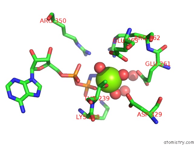
Mono view
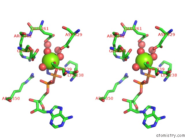
Stereo pair view
Mono view
Stereo pair view
A full contact list of Magnesium with other atoms in the Mg binding
site number 1 of Crystal Structure of Amp-Pnp Bound Enterococcus Hirae V1-Atpase [BV1] within 5.0Å range:
|
Magnesium binding site 2 out of 2 in 3vr6
Go back to
Magnesium binding site 2 out
of 2 in the Crystal Structure of Amp-Pnp Bound Enterococcus Hirae V1-Atpase [BV1]
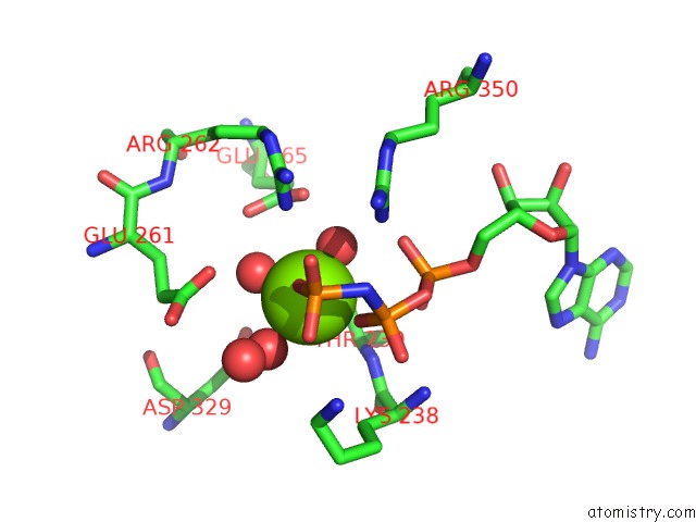
Mono view
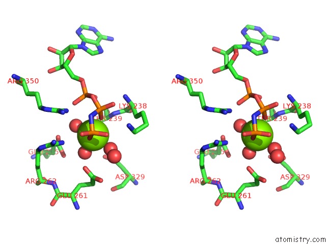
Stereo pair view
Mono view
Stereo pair view
A full contact list of Magnesium with other atoms in the Mg binding
site number 2 of Crystal Structure of Amp-Pnp Bound Enterococcus Hirae V1-Atpase [BV1] within 5.0Å range:
|
Reference:
S.Arai,
S.Saijo,
K.Suzuki,
K.Mizutani,
Y.Kakinuma,
Y.Ishizuka-Katsura,
N.Ohsawa,
T.Terada,
M.Shirouzu,
S.Yokoyama,
S.Iwata,
I.Yamato,
T.Murata.
Rotation Mechanism of Enterococcus Hirae V(1)-Atpase Based on Asymmetric Crystal Structures Nature V. 493 703 2013.
ISSN: ISSN 0028-0836
PubMed: 23334411
DOI: 10.1038/NATURE11778
Page generated: Mon Aug 11 04:40:57 2025
ISSN: ISSN 0028-0836
PubMed: 23334411
DOI: 10.1038/NATURE11778
Last articles
Mg in 6CA4Mg in 6C90
Mg in 6CA0
Mg in 6C9Y
Mg in 6C8Z
Mg in 6C8P
Mg in 6C8N
Mg in 6C8O
Mg in 6C8D
Mg in 6C8L