Magnesium »
PDB 3wdl-3wqc »
3wew »
Magnesium in PDB 3wew: Crystal Structure of Htdx (RV0241C) of Mycobacterium Tuberculosis at 2.4 A Resolution
Protein crystallography data
The structure of Crystal Structure of Htdx (RV0241C) of Mycobacterium Tuberculosis at 2.4 A Resolution, PDB code: 3wew
was solved by
R.Biswas,
D.Dutta,
A.K.Das,
with X-Ray Crystallography technique. A brief refinement statistics is given in the table below:
| Resolution Low / High (Å) | 19.49 / 2.40 |
| Space group | I 41 |
| Cell size a, b, c (Å), α, β, γ (°) | 61.504, 61.504, 143.803, 90.00, 90.00, 90.00 |
| R / Rfree (%) | 16.5 / 23 |
Magnesium Binding Sites:
The binding sites of Magnesium atom in the Crystal Structure of Htdx (RV0241C) of Mycobacterium Tuberculosis at 2.4 A Resolution
(pdb code 3wew). This binding sites where shown within
5.0 Angstroms radius around Magnesium atom.
In total only one binding site of Magnesium was determined in the Crystal Structure of Htdx (RV0241C) of Mycobacterium Tuberculosis at 2.4 A Resolution, PDB code: 3wew:
In total only one binding site of Magnesium was determined in the Crystal Structure of Htdx (RV0241C) of Mycobacterium Tuberculosis at 2.4 A Resolution, PDB code: 3wew:
Magnesium binding site 1 out of 1 in 3wew
Go back to
Magnesium binding site 1 out
of 1 in the Crystal Structure of Htdx (RV0241C) of Mycobacterium Tuberculosis at 2.4 A Resolution
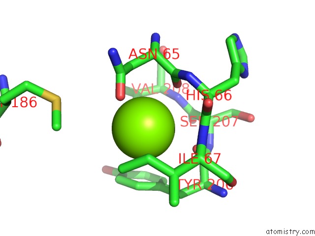
Mono view
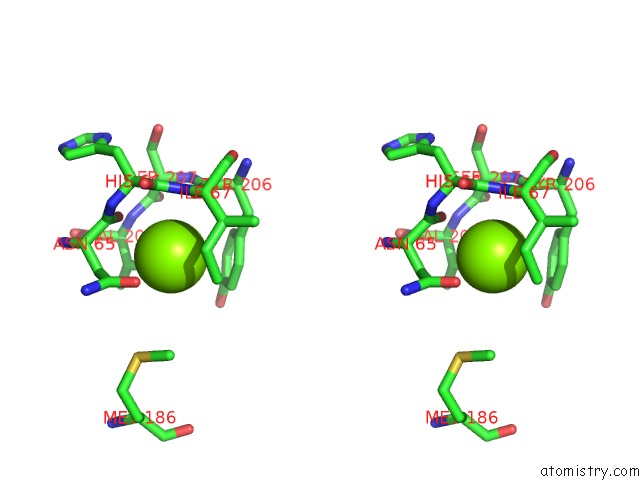
Stereo pair view
Mono view
Stereo pair view
A full contact list of Magnesium with other atoms in the Mg binding
site number 1 of Crystal Structure of Htdx (RV0241C) of Mycobacterium Tuberculosis at 2.4 A Resolution within 5.0Å range:
|
Reference:
R.Biswas,
D.Dutta,
A.K.Das.
Crystal Structure of A Putative Dehydratase Htdx of Mycobacterium Tuberculosis To Be Published.
Page generated: Thu Aug 15 13:23:55 2024
Last articles
Ca in 6HKZCa in 6HI0
Ca in 6HKJ
Ca in 6HK5
Ca in 6HCS
Ca in 6HHZ
Ca in 6HHY
Ca in 6HGM
Ca in 6HGB
Ca in 6HHM