Magnesium »
PDB 3zx5-4a2b »
457d »
Magnesium in PDB 457d: Molecular and Crystal Structure of D(CGCGMO6AATTCGCG): N6- Methoxyadenosine/ Thymidine Base-Pairs in B-Dna
Protein crystallography data
The structure of Molecular and Crystal Structure of D(CGCGMO6AATTCGCG): N6- Methoxyadenosine/ Thymidine Base-Pairs in B-Dna, PDB code: 457d
was solved by
T.Chatake,
A.Ono,
Y.Ueno,
A.Matsuda,
A.Takenaka,
with X-Ray Crystallography technique. A brief refinement statistics is given in the table below:
| Resolution Low / High (Å) | 5.00 / 2.00 |
| Space group | P 21 21 21 |
| Cell size a, b, c (Å), α, β, γ (°) | 24.900, 39.500, 66.200, 90.00, 90.00, 90.00 |
| R / Rfree (%) | 23.2 / 28.5 |
Magnesium Binding Sites:
The binding sites of Magnesium atom in the Molecular and Crystal Structure of D(CGCGMO6AATTCGCG): N6- Methoxyadenosine/ Thymidine Base-Pairs in B-Dna
(pdb code 457d). This binding sites where shown within
5.0 Angstroms radius around Magnesium atom.
In total only one binding site of Magnesium was determined in the Molecular and Crystal Structure of D(CGCGMO6AATTCGCG): N6- Methoxyadenosine/ Thymidine Base-Pairs in B-Dna, PDB code: 457d:
In total only one binding site of Magnesium was determined in the Molecular and Crystal Structure of D(CGCGMO6AATTCGCG): N6- Methoxyadenosine/ Thymidine Base-Pairs in B-Dna, PDB code: 457d:
Magnesium binding site 1 out of 1 in 457d
Go back to
Magnesium binding site 1 out
of 1 in the Molecular and Crystal Structure of D(CGCGMO6AATTCGCG): N6- Methoxyadenosine/ Thymidine Base-Pairs in B-Dna
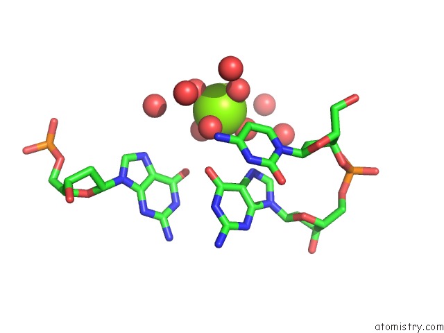
Mono view
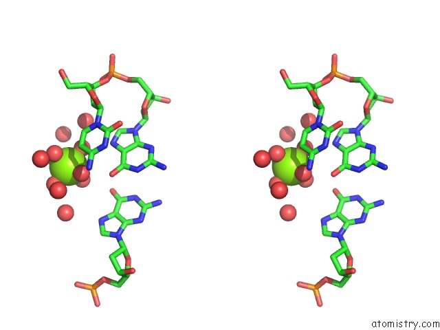
Stereo pair view
Mono view
Stereo pair view
A full contact list of Magnesium with other atoms in the Mg binding
site number 1 of Molecular and Crystal Structure of D(CGCGMO6AATTCGCG): N6- Methoxyadenosine/ Thymidine Base-Pairs in B-Dna within 5.0Å range:
|
Reference:
T.Chatake,
T.Hikima,
A.Ono,
Y.Ueno,
A.Matsuda,
A.Takenaka.
Crystallographic Studies on Damaged Dnas. II. N(6)-Methoxyadenine Can Present Two Alternate Faces For Watson-Crick Base-Pairing, Leading to Pyrimidine Transition Mutagenesis. J.Mol.Biol. V. 294 1223 1999.
ISSN: ISSN 0022-2836
PubMed: 10600380
DOI: 10.1006/JMBI.1999.3304
Page generated: Mon Aug 11 05:29:26 2025
ISSN: ISSN 0022-2836
PubMed: 10600380
DOI: 10.1006/JMBI.1999.3304
Last articles
Mg in 4M30Mg in 4M2Z
Mg in 4M32
Mg in 4LY6
Mg in 4M2A
Mg in 4M22
Mg in 4M0L
Mg in 4M1W
Mg in 4M1K
Mg in 4M0N