Magnesium »
PDB 4an2-4ava »
4as8 »
Magnesium in PDB 4as8: X-Ray Structure of the Cyan Fluorescent Protein Cerulean Cryoprotected with Ethylene Glycol
Protein crystallography data
The structure of X-Ray Structure of the Cyan Fluorescent Protein Cerulean Cryoprotected with Ethylene Glycol, PDB code: 4as8
was solved by
D.Von Stetten,
G.Batot,
M.Noirclerc-Savoye,
A.Royant,
with X-Ray Crystallography technique. A brief refinement statistics is given in the table below:
| Resolution Low / High (Å) | 46.75 / 1.02 |
| Space group | P 21 21 21 |
| Cell size a, b, c (Å), α, β, γ (°) | 51.344, 62.662, 70.205, 90.00, 90.00, 90.00 |
| R / Rfree (%) | 11.6 / 13.2 |
Magnesium Binding Sites:
The binding sites of Magnesium atom in the X-Ray Structure of the Cyan Fluorescent Protein Cerulean Cryoprotected with Ethylene Glycol
(pdb code 4as8). This binding sites where shown within
5.0 Angstroms radius around Magnesium atom.
In total only one binding site of Magnesium was determined in the X-Ray Structure of the Cyan Fluorescent Protein Cerulean Cryoprotected with Ethylene Glycol, PDB code: 4as8:
In total only one binding site of Magnesium was determined in the X-Ray Structure of the Cyan Fluorescent Protein Cerulean Cryoprotected with Ethylene Glycol, PDB code: 4as8:
Magnesium binding site 1 out of 1 in 4as8
Go back to
Magnesium binding site 1 out
of 1 in the X-Ray Structure of the Cyan Fluorescent Protein Cerulean Cryoprotected with Ethylene Glycol
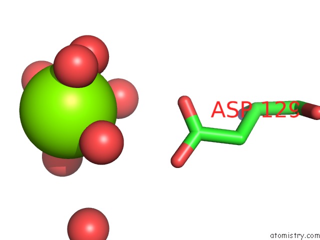
Mono view
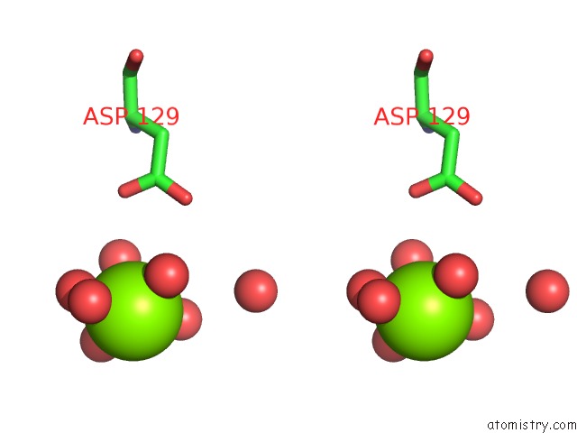
Stereo pair view
Mono view
Stereo pair view
A full contact list of Magnesium with other atoms in the Mg binding
site number 1 of X-Ray Structure of the Cyan Fluorescent Protein Cerulean Cryoprotected with Ethylene Glycol within 5.0Å range:
|
Reference:
D.Von Stetten,
G.Batot,
M.Noirclerc-Savoye,
A.Royant.
Alteration of Fluorescent Protein Spectroscopic Properties Upon Cryoprotection Acta Crystallogr.,Sect.D V. 68 1578 2012.
ISSN: ISSN 0907-4449
PubMed: 23090407
DOI: 10.1107/S0907444912037900
Page generated: Mon Aug 11 05:50:35 2025
ISSN: ISSN 0907-4449
PubMed: 23090407
DOI: 10.1107/S0907444912037900
Last articles
Mg in 6OD9Mg in 6OBJ
Mg in 6OE3
Mg in 6OB3
Mg in 6OAP
Mg in 6OAQ
Mg in 6OB2
Mg in 6O9P
Mg in 6O9O
Mg in 6O8E