Magnesium »
PDB 4fk1-4fs5 »
4fkx »
Magnesium in PDB 4fkx: Crystal Structure of Nucleoside Diphosphate Kinase B From Trypanosoma Brucei Bound to Cdp
Enzymatic activity of Crystal Structure of Nucleoside Diphosphate Kinase B From Trypanosoma Brucei Bound to Cdp
All present enzymatic activity of Crystal Structure of Nucleoside Diphosphate Kinase B From Trypanosoma Brucei Bound to Cdp:
2.7.4.6;
2.7.4.6;
Protein crystallography data
The structure of Crystal Structure of Nucleoside Diphosphate Kinase B From Trypanosoma Brucei Bound to Cdp, PDB code: 4fkx
was solved by
Seattle Structural Genomics Center For Infectious Disease (Ssgcid),
with X-Ray Crystallography technique. A brief refinement statistics is given in the table below:
| Resolution Low / High (Å) | 41.33 / 1.70 |
| Space group | C 2 2 21 |
| Cell size a, b, c (Å), α, β, γ (°) | 71.630, 121.370, 112.870, 90.00, 90.00, 90.00 |
| R / Rfree (%) | 15.2 / 18.2 |
Magnesium Binding Sites:
The binding sites of Magnesium atom in the Crystal Structure of Nucleoside Diphosphate Kinase B From Trypanosoma Brucei Bound to Cdp
(pdb code 4fkx). This binding sites where shown within
5.0 Angstroms radius around Magnesium atom.
In total 3 binding sites of Magnesium where determined in the Crystal Structure of Nucleoside Diphosphate Kinase B From Trypanosoma Brucei Bound to Cdp, PDB code: 4fkx:
Jump to Magnesium binding site number: 1; 2; 3;
In total 3 binding sites of Magnesium where determined in the Crystal Structure of Nucleoside Diphosphate Kinase B From Trypanosoma Brucei Bound to Cdp, PDB code: 4fkx:
Jump to Magnesium binding site number: 1; 2; 3;
Magnesium binding site 1 out of 3 in 4fkx
Go back to
Magnesium binding site 1 out
of 3 in the Crystal Structure of Nucleoside Diphosphate Kinase B From Trypanosoma Brucei Bound to Cdp
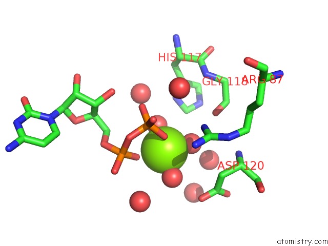
Mono view

Stereo pair view
Mono view
Stereo pair view
A full contact list of Magnesium with other atoms in the Mg binding
site number 1 of Crystal Structure of Nucleoside Diphosphate Kinase B From Trypanosoma Brucei Bound to Cdp within 5.0Å range:
|
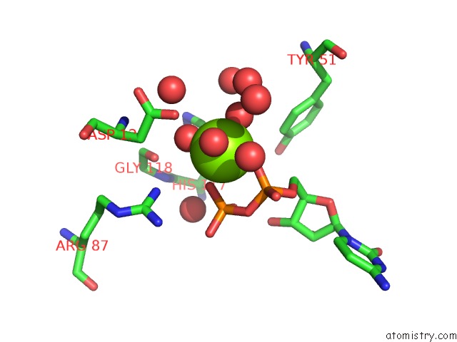
Magnesium binding site 2 out of 3 in 4fkx
Go back to
Magnesium binding site 2 out
of 3 in the Crystal Structure of Nucleoside Diphosphate Kinase B From Trypanosoma Brucei Bound to Cdp

Mono view

Stereo pair view
Mono view
Stereo pair view
A full contact list of Magnesium with other atoms in the Mg binding
site number 2 of Crystal Structure of Nucleoside Diphosphate Kinase B From Trypanosoma Brucei Bound to Cdp within 5.0Å range:
|
Magnesium binding site 3 out of 3 in 4fkx
Go back to
Magnesium binding site 3 out
of 3 in the Crystal Structure of Nucleoside Diphosphate Kinase B From Trypanosoma Brucei Bound to Cdp
Mono view
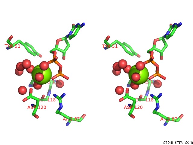
Stereo pair view
Mono view
Stereo pair view
A full contact list of Magnesium with other atoms in the Mg binding
site number 3 of Crystal Structure of Nucleoside Diphosphate Kinase B From Trypanosoma Brucei Bound to Cdp within 5.0Å range:
|
Reference:
A.S.Gardberg,
T.E.Edwards,
Seattle Structural Genomics Center For Infectious Disease(Ssgcid).
Crystal Structure of Nucleoside Diphosphate Kinase B From Trypanosoma Brucei Bound to Cdp To Be Published.
Page generated: Fri Aug 16 15:09:11 2024
Last articles
Fe in 2YXOFe in 2YRS
Fe in 2YXC
Fe in 2YNM
Fe in 2YVJ
Fe in 2YP1
Fe in 2YU2
Fe in 2YU1
Fe in 2YQB
Fe in 2YOO