Magnesium »
PDB 4fk1-4fs5 »
4fpv »
Magnesium in PDB 4fpv: Crystal Structure of D. Rerio TDP2 Complexed with Single Strand Dna Product
Protein crystallography data
The structure of Crystal Structure of D. Rerio TDP2 Complexed with Single Strand Dna Product, PDB code: 4fpv
was solved by
K.Shi,
K.Kurahashi,
H.Aihara,
with X-Ray Crystallography technique. A brief refinement statistics is given in the table below:
| Resolution Low / High (Å) | 35.15 / 1.73 |
| Space group | P 21 21 21 |
| Cell size a, b, c (Å), α, β, γ (°) | 58.070, 95.021, 104.506, 90.00, 90.00, 90.00 |
| R / Rfree (%) | 15.9 / 20.2 |
Other elements in 4fpv:
The structure of Crystal Structure of D. Rerio TDP2 Complexed with Single Strand Dna Product also contains other interesting chemical elements:
| Sodium | (Na) | 2 atoms |
Magnesium Binding Sites:
The binding sites of Magnesium atom in the Crystal Structure of D. Rerio TDP2 Complexed with Single Strand Dna Product
(pdb code 4fpv). This binding sites where shown within
5.0 Angstroms radius around Magnesium atom.
In total 2 binding sites of Magnesium where determined in the Crystal Structure of D. Rerio TDP2 Complexed with Single Strand Dna Product, PDB code: 4fpv:
Jump to Magnesium binding site number: 1; 2;
In total 2 binding sites of Magnesium where determined in the Crystal Structure of D. Rerio TDP2 Complexed with Single Strand Dna Product, PDB code: 4fpv:
Jump to Magnesium binding site number: 1; 2;
Magnesium binding site 1 out of 2 in 4fpv
Go back to
Magnesium binding site 1 out
of 2 in the Crystal Structure of D. Rerio TDP2 Complexed with Single Strand Dna Product
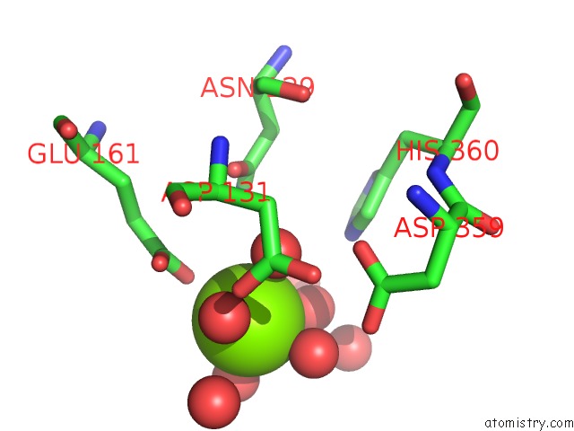
Mono view
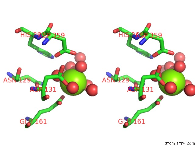
Stereo pair view
Mono view
Stereo pair view
A full contact list of Magnesium with other atoms in the Mg binding
site number 1 of Crystal Structure of D. Rerio TDP2 Complexed with Single Strand Dna Product within 5.0Å range:
|
Magnesium binding site 2 out of 2 in 4fpv
Go back to
Magnesium binding site 2 out
of 2 in the Crystal Structure of D. Rerio TDP2 Complexed with Single Strand Dna Product
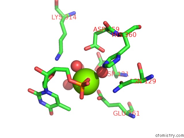
Mono view
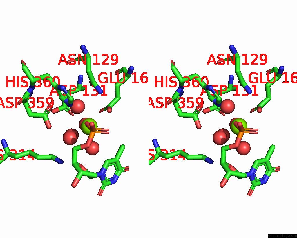
Stereo pair view
Mono view
Stereo pair view
A full contact list of Magnesium with other atoms in the Mg binding
site number 2 of Crystal Structure of D. Rerio TDP2 Complexed with Single Strand Dna Product within 5.0Å range:
|
Reference:
K.Shi,
K.Kurahashi,
R.Gao,
S.E.Tsutakawa,
J.A.Tainer,
Y.Pommier,
H.Aihara.
Structural Basis For Recognition of 5'-Phosphotyrosine Adducts By TDP2. Nat.Struct.Mol.Biol. V. 19 1372 2012.
ISSN: ISSN 1545-9993
PubMed: 23104058
DOI: 10.1038/NSMB.2423
Page generated: Fri Aug 16 15:14:31 2024
ISSN: ISSN 1545-9993
PubMed: 23104058
DOI: 10.1038/NSMB.2423
Last articles
Fe in 2YXOFe in 2YRS
Fe in 2YXC
Fe in 2YNM
Fe in 2YVJ
Fe in 2YP1
Fe in 2YU2
Fe in 2YU1
Fe in 2YQB
Fe in 2YOO