Magnesium »
PDB 4ix4-4j9k »
4izj »
Magnesium in PDB 4izj: Crystal Structure of Yellowtail Ascites Virus VP4 Protease with A Wild-Type Active Site Reveals Acyl-Enzyme Complexes and Product Complexes.
Enzymatic activity of Crystal Structure of Yellowtail Ascites Virus VP4 Protease with A Wild-Type Active Site Reveals Acyl-Enzyme Complexes and Product Complexes.
All present enzymatic activity of Crystal Structure of Yellowtail Ascites Virus VP4 Protease with A Wild-Type Active Site Reveals Acyl-Enzyme Complexes and Product Complexes.:
3.4.21.115;
3.4.21.115;
Protein crystallography data
The structure of Crystal Structure of Yellowtail Ascites Virus VP4 Protease with A Wild-Type Active Site Reveals Acyl-Enzyme Complexes and Product Complexes., PDB code: 4izj
was solved by
M.Paetzel,
I.Y.W.Chung,
with X-Ray Crystallography technique. A brief refinement statistics is given in the table below:
| Resolution Low / High (Å) | 64.30 / 2.50 |
| Space group | P 1 21 1 |
| Cell size a, b, c (Å), α, β, γ (°) | 41.610, 64.330, 187.740, 90.00, 95.80, 90.00 |
| R / Rfree (%) | 17.8 / 24.4 |
Magnesium Binding Sites:
The binding sites of Magnesium atom in the Crystal Structure of Yellowtail Ascites Virus VP4 Protease with A Wild-Type Active Site Reveals Acyl-Enzyme Complexes and Product Complexes.
(pdb code 4izj). This binding sites where shown within
5.0 Angstroms radius around Magnesium atom.
In total only one binding site of Magnesium was determined in the Crystal Structure of Yellowtail Ascites Virus VP4 Protease with A Wild-Type Active Site Reveals Acyl-Enzyme Complexes and Product Complexes., PDB code: 4izj:
In total only one binding site of Magnesium was determined in the Crystal Structure of Yellowtail Ascites Virus VP4 Protease with A Wild-Type Active Site Reveals Acyl-Enzyme Complexes and Product Complexes., PDB code: 4izj:
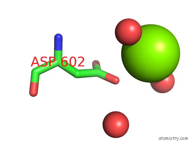
Magnesium binding site 1 out of 1 in 4izj
Go back to
Magnesium binding site 1 out
of 1 in the Crystal Structure of Yellowtail Ascites Virus VP4 Protease with A Wild-Type Active Site Reveals Acyl-Enzyme Complexes and Product Complexes.
Mono view

Stereo pair view
Mono view
Stereo pair view
A full contact list of Magnesium with other atoms in the Mg binding
site number 1 of Crystal Structure of Yellowtail Ascites Virus VP4 Protease with A Wild-Type Active Site Reveals Acyl-Enzyme Complexes and Product Complexes. within 5.0Å range:
|
Reference:
I.Y.Chung,
M.Paetzel.
Crystal Structures of Yellowtail Ascites Virus VP4 Protease: Trapping An Internal Cleavage Site Trans Acyl-Enzyme Complex in A Native Ser/Lys Dyad Active Site. J.Biol.Chem. V. 288 13068 2013.
ISSN: ISSN 0021-9258
PubMed: 23511637
DOI: 10.1074/JBC.M112.386953
Page generated: Mon Aug 11 14:30:05 2025
ISSN: ISSN 0021-9258
PubMed: 23511637
DOI: 10.1074/JBC.M112.386953
Last articles
Mg in 4JJSMg in 4JJ2
Mg in 4JIW
Mg in 4JIV
Mg in 4JIB
Mg in 4JI4
Mg in 4JI5
Mg in 4JI1
Mg in 4JI0
Mg in 4JI2