Magnesium »
PDB 4kvy-4la6 »
4kws »
Magnesium in PDB 4kws: Crystal Structure of D-Mannonate Dehydratase From Chromohalobacter Salexigens Complexed with Mg and Glycerol
Enzymatic activity of Crystal Structure of D-Mannonate Dehydratase From Chromohalobacter Salexigens Complexed with Mg and Glycerol
All present enzymatic activity of Crystal Structure of D-Mannonate Dehydratase From Chromohalobacter Salexigens Complexed with Mg and Glycerol:
4.2.1.8;
4.2.1.8;
Protein crystallography data
The structure of Crystal Structure of D-Mannonate Dehydratase From Chromohalobacter Salexigens Complexed with Mg and Glycerol, PDB code: 4kws
was solved by
A.A.Fedorov,
E.V.Fedorov,
D.Wichelecki,
J.A.Gerlt,
S.C.Almo,
with X-Ray Crystallography technique. A brief refinement statistics is given in the table below:
| Resolution Low / High (Å) | 40.05 / 1.64 |
| Space group | C 1 2 1 |
| Cell size a, b, c (Å), α, β, γ (°) | 195.273, 85.762, 195.097, 90.00, 110.31, 90.00 |
| R / Rfree (%) | 13.9 / 16.3 |
Other elements in 4kws:
The structure of Crystal Structure of D-Mannonate Dehydratase From Chromohalobacter Salexigens Complexed with Mg and Glycerol also contains other interesting chemical elements:
| Chlorine | (Cl) | 4 atoms |
Magnesium Binding Sites:
Pages:
>>> Page 1 <<< Page 2, Binding sites: 11 - 13;Binding sites:
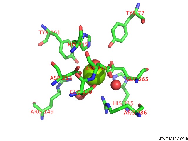
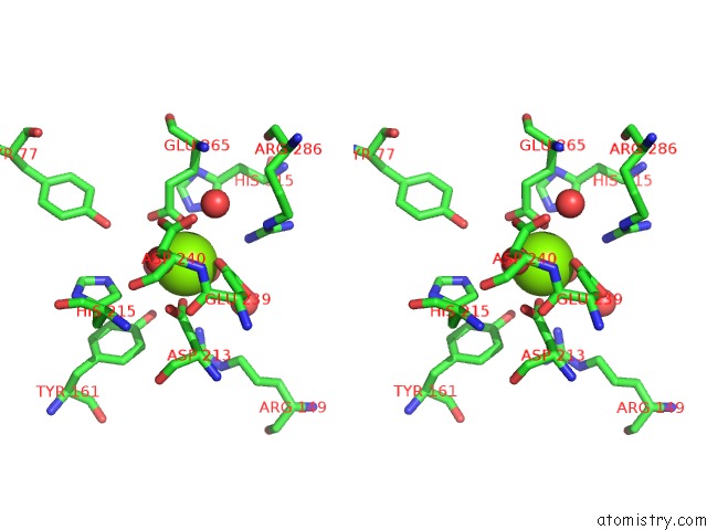
The binding sites of Magnesium atom in the Crystal Structure of D-Mannonate Dehydratase From Chromohalobacter Salexigens Complexed with Mg and Glycerol (pdb code 4kws). This binding sites where shown within 5.0 Angstroms radius around Magnesium atom.In total 13 binding sites of Magnesium where determined in the Crystal Structure of D-Mannonate Dehydratase From Chromohalobacter Salexigens Complexed with Mg and Glycerol, PDB code: 4kws:
Jump to Magnesium binding site number: 1; 2; 3; 4; 5; 6; 7; 8; 9; 10;
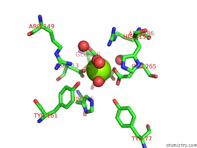
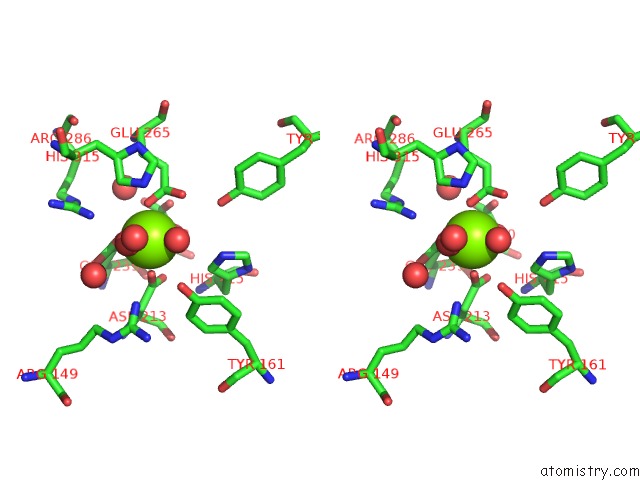
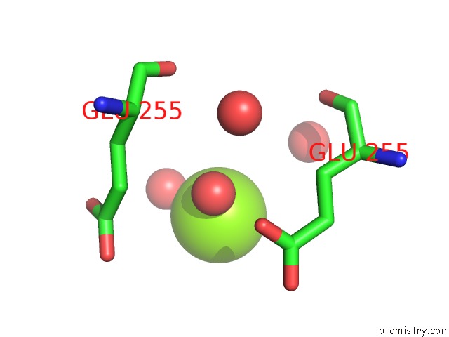
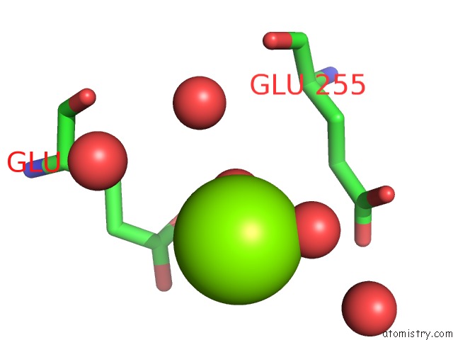
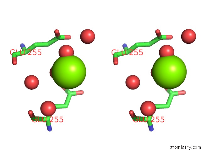
Magnesium binding site 1 out of 13 in 4kws
Go back to
Magnesium binding site 1 out
of 13 in the Crystal Structure of D-Mannonate Dehydratase From Chromohalobacter Salexigens Complexed with Mg and Glycerol

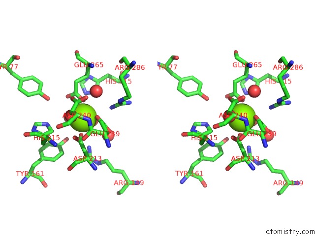
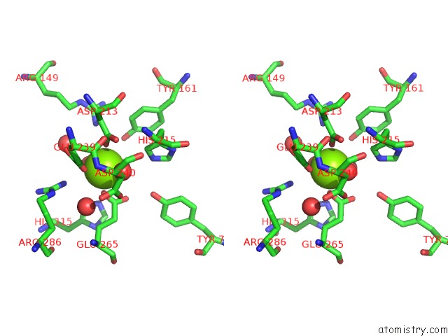
Mono view
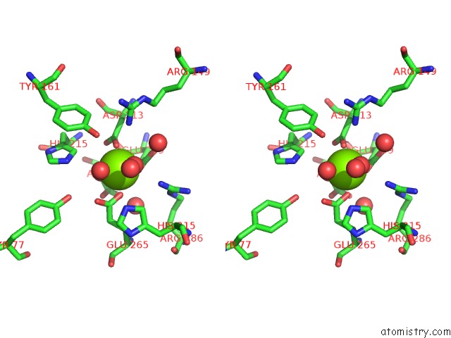
Stereo pair view
Mono view
Stereo pair view
A full contact list of Magnesium with other atoms in the Mg binding
site number 1 of Crystal Structure of D-Mannonate Dehydratase From Chromohalobacter Salexigens Complexed with Mg and Glycerol within 5.0Å range:
|
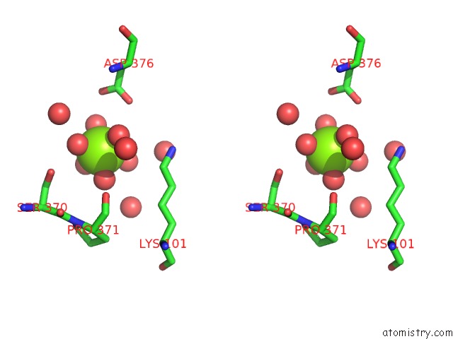
Magnesium binding site 2 out of 13 in 4kws
Go back to
Magnesium binding site 2 out
of 13 in the Crystal Structure of D-Mannonate Dehydratase From Chromohalobacter Salexigens Complexed with Mg and Glycerol

Mono view
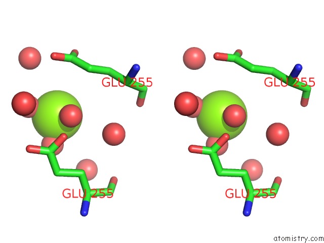
Stereo pair view
Mono view
Stereo pair view
A full contact list of Magnesium with other atoms in the Mg binding
site number 2 of Crystal Structure of D-Mannonate Dehydratase From Chromohalobacter Salexigens Complexed with Mg and Glycerol within 5.0Å range:
|
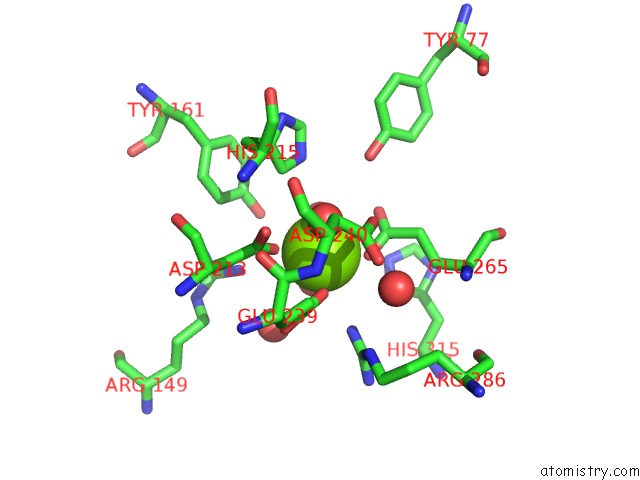
Magnesium binding site 3 out of 13 in 4kws
Go back to
Magnesium binding site 3 out
of 13 in the Crystal Structure of D-Mannonate Dehydratase From Chromohalobacter Salexigens Complexed with Mg and Glycerol
Mono view
Stereo pair view
Mono view
Stereo pair view
A full contact list of Magnesium with other atoms in the Mg binding
site number 3 of Crystal Structure of D-Mannonate Dehydratase From Chromohalobacter Salexigens Complexed with Mg and Glycerol within 5.0Å range:
|
Magnesium binding site 4 out of 13 in 4kws
Go back to
Magnesium binding site 4 out
of 13 in the Crystal Structure of D-Mannonate Dehydratase From Chromohalobacter Salexigens Complexed with Mg and Glycerol
Mono view

Stereo pair view
Mono view
Stereo pair view
A full contact list of Magnesium with other atoms in the Mg binding
site number 4 of Crystal Structure of D-Mannonate Dehydratase From Chromohalobacter Salexigens Complexed with Mg and Glycerol within 5.0Å range:
|
Magnesium binding site 5 out of 13 in 4kws
Go back to
Magnesium binding site 5 out
of 13 in the Crystal Structure of D-Mannonate Dehydratase From Chromohalobacter Salexigens Complexed with Mg and Glycerol
Mono view
Stereo pair view
Mono view
Stereo pair view
A full contact list of Magnesium with other atoms in the Mg binding
site number 5 of Crystal Structure of D-Mannonate Dehydratase From Chromohalobacter Salexigens Complexed with Mg and Glycerol within 5.0Å range:
|
Magnesium binding site 6 out of 13 in 4kws
Go back to
Magnesium binding site 6 out
of 13 in the Crystal Structure of D-Mannonate Dehydratase From Chromohalobacter Salexigens Complexed with Mg and Glycerol

Mono view

Stereo pair view
Mono view
Stereo pair view
A full contact list of Magnesium with other atoms in the Mg binding
site number 6 of Crystal Structure of D-Mannonate Dehydratase From Chromohalobacter Salexigens Complexed with Mg and Glycerol within 5.0Å range:
|
Magnesium binding site 7 out of 13 in 4kws
Go back to
Magnesium binding site 7 out
of 13 in the Crystal Structure of D-Mannonate Dehydratase From Chromohalobacter Salexigens Complexed with Mg and Glycerol

Mono view
Stereo pair view
Mono view
Stereo pair view
A full contact list of Magnesium with other atoms in the Mg binding
site number 7 of Crystal Structure of D-Mannonate Dehydratase From Chromohalobacter Salexigens Complexed with Mg and Glycerol within 5.0Å range:
|
Magnesium binding site 8 out of 13 in 4kws
Go back to
Magnesium binding site 8 out
of 13 in the Crystal Structure of D-Mannonate Dehydratase From Chromohalobacter Salexigens Complexed with Mg and Glycerol
Mono view
Stereo pair view
Mono view
Stereo pair view
A full contact list of Magnesium with other atoms in the Mg binding
site number 8 of Crystal Structure of D-Mannonate Dehydratase From Chromohalobacter Salexigens Complexed with Mg and Glycerol within 5.0Å range:
|
Magnesium binding site 9 out of 13 in 4kws
Go back to
Magnesium binding site 9 out
of 13 in the Crystal Structure of D-Mannonate Dehydratase From Chromohalobacter Salexigens Complexed with Mg and Glycerol

Mono view
Stereo pair view
Mono view
Stereo pair view
A full contact list of Magnesium with other atoms in the Mg binding
site number 9 of Crystal Structure of D-Mannonate Dehydratase From Chromohalobacter Salexigens Complexed with Mg and Glycerol within 5.0Å range:
|
Magnesium binding site 10 out of 13 in 4kws
Go back to
Magnesium binding site 10 out
of 13 in the Crystal Structure of D-Mannonate Dehydratase From Chromohalobacter Salexigens Complexed with Mg and Glycerol
Mono view
Stereo pair view
Mono view
Stereo pair view
A full contact list of Magnesium with other atoms in the Mg binding
site number 10 of Crystal Structure of D-Mannonate Dehydratase From Chromohalobacter Salexigens Complexed with Mg and Glycerol within 5.0Å range:
|
Reference:
A.A.Fedorov,
E.V.Fedorov,
D.Wichelecki,
J.A.Gerlt,
S.C.Almo.
Crystal Structure of D-Mannonate Dehydratase From Chromohalobacter Salexigens Complexed with Mg and Glycerol To Be Published.
Page generated: Mon Aug 11 18:08:00 2025
Last articles
Mn in 4M8DMn in 4MIG
Mn in 4MK5
Mn in 4MK1
Mn in 4MK2
Mn in 4M5R
Mn in 4MDA
Mn in 4MGH
Mn in 4M5U
Mn in 4M3C