Magnesium »
PDB 4lja-4lsh »
4lno »
Magnesium in PDB 4lno: B. Subtilis Glutamine Synthetase Structures Reveal Large Active Site Conformational Changes and Basis For Isoenzyme Specific Regulation: Form Two of Gs-1
Enzymatic activity of B. Subtilis Glutamine Synthetase Structures Reveal Large Active Site Conformational Changes and Basis For Isoenzyme Specific Regulation: Form Two of Gs-1
All present enzymatic activity of B. Subtilis Glutamine Synthetase Structures Reveal Large Active Site Conformational Changes and Basis For Isoenzyme Specific Regulation: Form Two of Gs-1:
6.3.1.2;
6.3.1.2;
Protein crystallography data
The structure of B. Subtilis Glutamine Synthetase Structures Reveal Large Active Site Conformational Changes and Basis For Isoenzyme Specific Regulation: Form Two of Gs-1, PDB code: 4lno
was solved by
M.A.Schumacher,
N.Chinnam,
N.Tonthat,
S.Fisher,
L.Wray,
with X-Ray Crystallography technique. A brief refinement statistics is given in the table below:
| Resolution Low / High (Å) | 71.68 / 2.90 |
| Space group | C 1 2 1 |
| Cell size a, b, c (Å), α, β, γ (°) | 209.860, 138.940, 144.730, 90.00, 125.17, 90.00 |
| R / Rfree (%) | 19.6 / 25.6 |
Magnesium Binding Sites:
Pages:
>>> Page 1 <<< Page 2, Binding sites: 11 - 12;Binding sites:
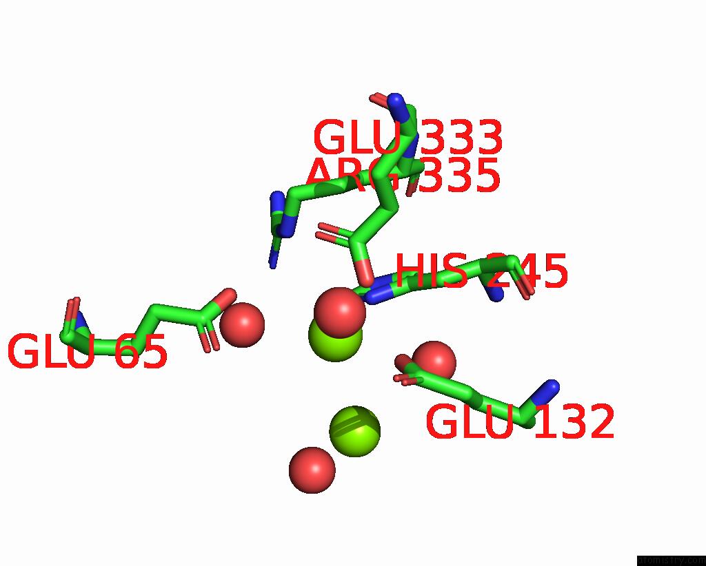
The binding sites of Magnesium atom in the B. Subtilis Glutamine Synthetase Structures Reveal Large Active Site Conformational Changes and Basis For Isoenzyme Specific Regulation: Form Two of Gs-1 (pdb code 4lno). This binding sites where shown within 5.0 Angstroms radius around Magnesium atom.In total 12 binding sites of Magnesium where determined in the B. Subtilis Glutamine Synthetase Structures Reveal Large Active Site Conformational Changes and Basis For Isoenzyme Specific Regulation: Form Two of Gs-1, PDB code: 4lno:
Jump to Magnesium binding site number: 1; 2; 3; 4; 5; 6; 7; 8; 9; 10;
Magnesium binding site 1 out of 12 in 4lno
Go back to
Magnesium binding site 1 out
of 12 in the B. Subtilis Glutamine Synthetase Structures Reveal Large Active Site Conformational Changes and Basis For Isoenzyme Specific Regulation: Form Two of Gs-1

Mono view

Stereo pair view
Mono view
Stereo pair view
A full contact list of Magnesium with other atoms in the Mg binding
site number 1 of B. Subtilis Glutamine Synthetase Structures Reveal Large Active Site Conformational Changes and Basis For Isoenzyme Specific Regulation: Form Two of Gs-1 within 5.0Å range:
|
Magnesium binding site 2 out of 12 in 4lno
Go back to
Magnesium binding site 2 out
of 12 in the B. Subtilis Glutamine Synthetase Structures Reveal Large Active Site Conformational Changes and Basis For Isoenzyme Specific Regulation: Form Two of Gs-1

Mono view

Stereo pair view
Mono view
Stereo pair view
A full contact list of Magnesium with other atoms in the Mg binding
site number 2 of B. Subtilis Glutamine Synthetase Structures Reveal Large Active Site Conformational Changes and Basis For Isoenzyme Specific Regulation: Form Two of Gs-1 within 5.0Å range:
|
Magnesium binding site 3 out of 12 in 4lno
Go back to
Magnesium binding site 3 out
of 12 in the B. Subtilis Glutamine Synthetase Structures Reveal Large Active Site Conformational Changes and Basis For Isoenzyme Specific Regulation: Form Two of Gs-1

Mono view

Stereo pair view
Mono view
Stereo pair view
A full contact list of Magnesium with other atoms in the Mg binding
site number 3 of B. Subtilis Glutamine Synthetase Structures Reveal Large Active Site Conformational Changes and Basis For Isoenzyme Specific Regulation: Form Two of Gs-1 within 5.0Å range:
|
Magnesium binding site 4 out of 12 in 4lno
Go back to
Magnesium binding site 4 out
of 12 in the B. Subtilis Glutamine Synthetase Structures Reveal Large Active Site Conformational Changes and Basis For Isoenzyme Specific Regulation: Form Two of Gs-1
Mono view

Stereo pair view
Mono view
Stereo pair view
A full contact list of Magnesium with other atoms in the Mg binding
site number 4 of B. Subtilis Glutamine Synthetase Structures Reveal Large Active Site Conformational Changes and Basis For Isoenzyme Specific Regulation: Form Two of Gs-1 within 5.0Å range:
|
Magnesium binding site 5 out of 12 in 4lno
Go back to
Magnesium binding site 5 out
of 12 in the B. Subtilis Glutamine Synthetase Structures Reveal Large Active Site Conformational Changes and Basis For Isoenzyme Specific Regulation: Form Two of Gs-1

Mono view

Stereo pair view
Mono view
Stereo pair view
A full contact list of Magnesium with other atoms in the Mg binding
site number 5 of B. Subtilis Glutamine Synthetase Structures Reveal Large Active Site Conformational Changes and Basis For Isoenzyme Specific Regulation: Form Two of Gs-1 within 5.0Å range:
|
Magnesium binding site 6 out of 12 in 4lno
Go back to
Magnesium binding site 6 out
of 12 in the B. Subtilis Glutamine Synthetase Structures Reveal Large Active Site Conformational Changes and Basis For Isoenzyme Specific Regulation: Form Two of Gs-1

Mono view

Stereo pair view
Mono view
Stereo pair view
A full contact list of Magnesium with other atoms in the Mg binding
site number 6 of B. Subtilis Glutamine Synthetase Structures Reveal Large Active Site Conformational Changes and Basis For Isoenzyme Specific Regulation: Form Two of Gs-1 within 5.0Å range:
|
Magnesium binding site 7 out of 12 in 4lno
Go back to
Magnesium binding site 7 out
of 12 in the B. Subtilis Glutamine Synthetase Structures Reveal Large Active Site Conformational Changes and Basis For Isoenzyme Specific Regulation: Form Two of Gs-1

Mono view

Stereo pair view
Mono view
Stereo pair view
A full contact list of Magnesium with other atoms in the Mg binding
site number 7 of B. Subtilis Glutamine Synthetase Structures Reveal Large Active Site Conformational Changes and Basis For Isoenzyme Specific Regulation: Form Two of Gs-1 within 5.0Å range:
|
Magnesium binding site 8 out of 12 in 4lno
Go back to
Magnesium binding site 8 out
of 12 in the B. Subtilis Glutamine Synthetase Structures Reveal Large Active Site Conformational Changes and Basis For Isoenzyme Specific Regulation: Form Two of Gs-1

Mono view

Stereo pair view
Mono view
Stereo pair view
A full contact list of Magnesium with other atoms in the Mg binding
site number 8 of B. Subtilis Glutamine Synthetase Structures Reveal Large Active Site Conformational Changes and Basis For Isoenzyme Specific Regulation: Form Two of Gs-1 within 5.0Å range:
|
Magnesium binding site 9 out of 12 in 4lno
Go back to
Magnesium binding site 9 out
of 12 in the B. Subtilis Glutamine Synthetase Structures Reveal Large Active Site Conformational Changes and Basis For Isoenzyme Specific Regulation: Form Two of Gs-1

Mono view

Stereo pair view
Mono view
Stereo pair view
A full contact list of Magnesium with other atoms in the Mg binding
site number 9 of B. Subtilis Glutamine Synthetase Structures Reveal Large Active Site Conformational Changes and Basis For Isoenzyme Specific Regulation: Form Two of Gs-1 within 5.0Å range:
|
Magnesium binding site 10 out of 12 in 4lno
Go back to
Magnesium binding site 10 out
of 12 in the B. Subtilis Glutamine Synthetase Structures Reveal Large Active Site Conformational Changes and Basis For Isoenzyme Specific Regulation: Form Two of Gs-1

Mono view

Stereo pair view
Mono view
Stereo pair view
A full contact list of Magnesium with other atoms in the Mg binding
site number 10 of B. Subtilis Glutamine Synthetase Structures Reveal Large Active Site Conformational Changes and Basis For Isoenzyme Specific Regulation: Form Two of Gs-1 within 5.0Å range:
|
Reference:
D.S.Murray,
N.Chinnam,
N.K.Tonthat,
T.Whitfill,
L.V.Wray,
S.H.Fisher,
M.A.Schumacher.
Structures of the Bacillus Subtilis Glutamine Synthetase Dodecamer Reveal Large Intersubunit Catalytic Conformational Changes Linked to A Unique Feedback Inhibition Mechanism. J.Biol.Chem. V. 288 35801 2013.
ISSN: ISSN 0021-9258
PubMed: 24158439
DOI: 10.1074/JBC.M113.519496
Page generated: Mon Aug 11 19:58:48 2025
ISSN: ISSN 0021-9258
PubMed: 24158439
DOI: 10.1074/JBC.M113.519496
Last articles
Mg in 4RIIMg in 4RIG
Mg in 4RIH
Mg in 4RI2
Mg in 4RHX
Mg in 4RHU
Mg in 4RHT
Mg in 4RHD
Mg in 4RH7
Mg in 4RET