Magnesium »
PDB 4mff-4mpo »
4mn0 »
Magnesium in PDB 4mn0: Spatial Structure of the Novel Light-Sensitive Photoprotein Berovin From the Ctenophore Beroe Abyssicola in the CA2+-Loaded Apoprotein Conformation State
Protein crystallography data
The structure of Spatial Structure of the Novel Light-Sensitive Photoprotein Berovin From the Ctenophore Beroe Abyssicola in the CA2+-Loaded Apoprotein Conformation State, PDB code: 4mn0
was solved by
Z.J.Liu,
G.A.Stepanyuk,
E.S.Vysotski,
J.Lee,
J.P.Rose,
B.C.Wang,
Southeastcollaboratory For Structural Genomics (Secsg),
with X-Ray Crystallography technique. A brief refinement statistics is given in the table below:
| Resolution Low / High (Å) | 40.78 / 1.90 |
| Space group | C 1 2 1 |
| Cell size a, b, c (Å), α, β, γ (°) | 101.770, 33.899, 77.406, 90.00, 126.74, 90.00 |
| R / Rfree (%) | 19.3 / 25.6 |
Other elements in 4mn0:
The structure of Spatial Structure of the Novel Light-Sensitive Photoprotein Berovin From the Ctenophore Beroe Abyssicola in the CA2+-Loaded Apoprotein Conformation State also contains other interesting chemical elements:
| Calcium | (Ca) | 3 atoms |
Magnesium Binding Sites:
The binding sites of Magnesium atom in the Spatial Structure of the Novel Light-Sensitive Photoprotein Berovin From the Ctenophore Beroe Abyssicola in the CA2+-Loaded Apoprotein Conformation State
(pdb code 4mn0). This binding sites where shown within
5.0 Angstroms radius around Magnesium atom.
In total only one binding site of Magnesium was determined in the Spatial Structure of the Novel Light-Sensitive Photoprotein Berovin From the Ctenophore Beroe Abyssicola in the CA2+-Loaded Apoprotein Conformation State, PDB code: 4mn0:
In total only one binding site of Magnesium was determined in the Spatial Structure of the Novel Light-Sensitive Photoprotein Berovin From the Ctenophore Beroe Abyssicola in the CA2+-Loaded Apoprotein Conformation State, PDB code: 4mn0:
Magnesium binding site 1 out of 1 in 4mn0
Go back to
Magnesium binding site 1 out
of 1 in the Spatial Structure of the Novel Light-Sensitive Photoprotein Berovin From the Ctenophore Beroe Abyssicola in the CA2+-Loaded Apoprotein Conformation State
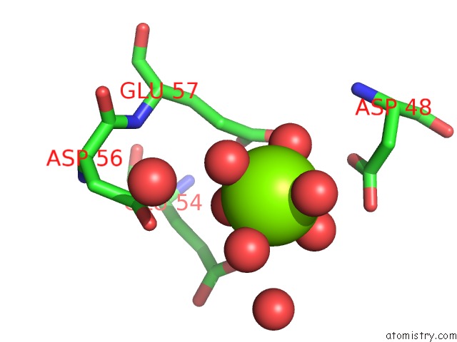
Mono view
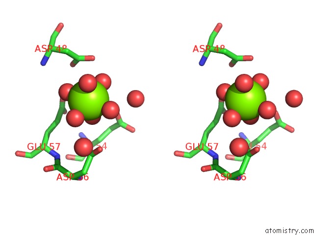
Stereo pair view
Mono view
Stereo pair view
A full contact list of Magnesium with other atoms in the Mg binding
site number 1 of Spatial Structure of the Novel Light-Sensitive Photoprotein Berovin From the Ctenophore Beroe Abyssicola in the CA2+-Loaded Apoprotein Conformation State within 5.0Å range:
|
Reference:
G.A.Stepanyuk,
Z.J.Liu,
L.P.Burakova,
J.Lee,
J.Rose,
E.S.Vysotski,
B.C.Wang.
Spatial Structure of the Novel Light-Sensitive Photoprotein Berovin From the Ctenophore Beroe Abyssicola in the Ca(2+)-Loaded Apoprotein Conformation State. Biochim.Biophys.Acta V.1834 2139 2013.
ISSN: ISSN 0006-3002
PubMed: 23891746
DOI: 10.1016/J.BBAPAP.2013.07.006
Page generated: Mon Aug 11 20:30:02 2025
ISSN: ISSN 0006-3002
PubMed: 23891746
DOI: 10.1016/J.BBAPAP.2013.07.006
Last articles
Mg in 4QLVMg in 4QOX
Mg in 4QN0
Mg in 4QNY
Mg in 4QLU
Mg in 4QNR
Mg in 4QMU
Mg in 4QML
Mg in 4QM7
Mg in 4QM6