Magnesium »
PDB 4mq9-4nb4 »
4myq »
Magnesium in PDB 4myq: Selective Inhibition of the Catalytic Domain of Human Phosphodiesterase 4B with A-33
Enzymatic activity of Selective Inhibition of the Catalytic Domain of Human Phosphodiesterase 4B with A-33
All present enzymatic activity of Selective Inhibition of the Catalytic Domain of Human Phosphodiesterase 4B with A-33:
3.1.4.17;
3.1.4.17;
Protein crystallography data
The structure of Selective Inhibition of the Catalytic Domain of Human Phosphodiesterase 4B with A-33, PDB code: 4myq
was solved by
D.Fox Iii,
T.E.Edwards,
with X-Ray Crystallography technique. A brief refinement statistics is given in the table below:
| Resolution Low / High (Å) | 49.85 / 1.90 |
| Space group | P 42 |
| Cell size a, b, c (Å), α, β, γ (°) | 99.700, 99.700, 47.020, 90.00, 90.00, 90.00 |
| R / Rfree (%) | 18.8 / 22.2 |
Other elements in 4myq:
The structure of Selective Inhibition of the Catalytic Domain of Human Phosphodiesterase 4B with A-33 also contains other interesting chemical elements:
| Zinc | (Zn) | 1 atom |
| Chlorine | (Cl) | 1 atom |
| Sodium | (Na) | 1 atom |
Magnesium Binding Sites:
The binding sites of Magnesium atom in the Selective Inhibition of the Catalytic Domain of Human Phosphodiesterase 4B with A-33
(pdb code 4myq). This binding sites where shown within
5.0 Angstroms radius around Magnesium atom.
In total only one binding site of Magnesium was determined in the Selective Inhibition of the Catalytic Domain of Human Phosphodiesterase 4B with A-33, PDB code: 4myq:
In total only one binding site of Magnesium was determined in the Selective Inhibition of the Catalytic Domain of Human Phosphodiesterase 4B with A-33, PDB code: 4myq:
Magnesium binding site 1 out of 1 in 4myq
Go back to
Magnesium binding site 1 out
of 1 in the Selective Inhibition of the Catalytic Domain of Human Phosphodiesterase 4B with A-33
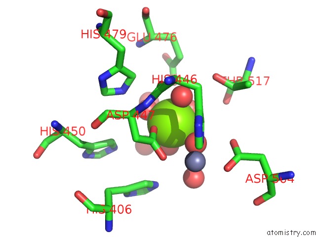
Mono view
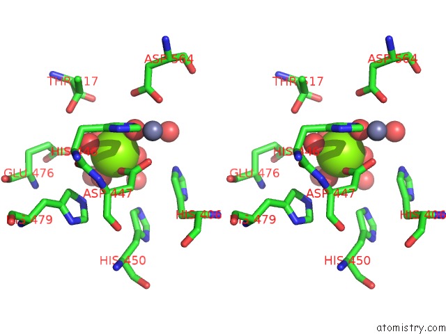
Stereo pair view
Mono view
Stereo pair view
A full contact list of Magnesium with other atoms in the Mg binding
site number 1 of Selective Inhibition of the Catalytic Domain of Human Phosphodiesterase 4B with A-33 within 5.0Å range:
|
Reference:
D.Fox,
A.B.Burgin,
M.E.Gurney.
Structural Basis For the Design of Selective Phosphodiesterase 4B Inhibitors. Cell Signal V. 26 657 2014.
PubMed: 24361374
DOI: 10.1016/J.CELLSIG.2013.12.003
Page generated: Mon Aug 11 20:34:05 2025
PubMed: 24361374
DOI: 10.1016/J.CELLSIG.2013.12.003
Last articles
Mg in 9MOMMg in 9MOL
Mg in 9MOK
Mg in 9MOI
Mg in 9MOD
Mg in 9MOC
Mg in 9MOB
Mg in 9MOA
Mg in 9MO8
Mg in 9MO9