Magnesium »
PDB 4osq-4p8r »
4p02 »
Magnesium in PDB 4p02: Structure of Bacterial Cellulose Synthase with Cyclic-Di-Gmp Bound.
Enzymatic activity of Structure of Bacterial Cellulose Synthase with Cyclic-Di-Gmp Bound.
All present enzymatic activity of Structure of Bacterial Cellulose Synthase with Cyclic-Di-Gmp Bound.:
2.4.1.12;
2.4.1.12;
Protein crystallography data
The structure of Structure of Bacterial Cellulose Synthase with Cyclic-Di-Gmp Bound., PDB code: 4p02
was solved by
J.L.W.Morgan,
J.T.Mcnamara,
J.Zimmer,
with X-Ray Crystallography technique. A brief refinement statistics is given in the table below:
| Resolution Low / High (Å) | 19.99 / 2.65 |
| Space group | P 21 21 21 |
| Cell size a, b, c (Å), α, β, γ (°) | 67.640, 214.660, 220.400, 90.00, 90.00, 90.00 |
| R / Rfree (%) | 19.9 / 23 |
Magnesium Binding Sites:
The binding sites of Magnesium atom in the Structure of Bacterial Cellulose Synthase with Cyclic-Di-Gmp Bound.
(pdb code 4p02). This binding sites where shown within
5.0 Angstroms radius around Magnesium atom.
In total only one binding site of Magnesium was determined in the Structure of Bacterial Cellulose Synthase with Cyclic-Di-Gmp Bound., PDB code: 4p02:
In total only one binding site of Magnesium was determined in the Structure of Bacterial Cellulose Synthase with Cyclic-Di-Gmp Bound., PDB code: 4p02:
Magnesium binding site 1 out of 1 in 4p02
Go back to
Magnesium binding site 1 out
of 1 in the Structure of Bacterial Cellulose Synthase with Cyclic-Di-Gmp Bound.

Mono view
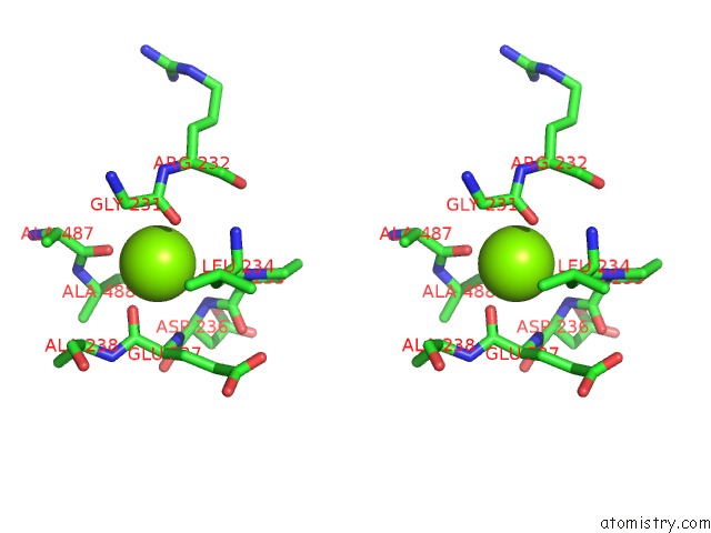
Stereo pair view
Mono view
Stereo pair view
A full contact list of Magnesium with other atoms in the Mg binding
site number 1 of Structure of Bacterial Cellulose Synthase with Cyclic-Di-Gmp Bound. within 5.0Å range:
|
Reference:
J.L.Morgan,
J.T.Mcnamara,
J.Zimmer.
Mechanism of Activation of Bacterial Cellulose Synthase By Cyclic Di-Gmp. Nat.Struct.Mol.Biol. V. 21 489 2014.
ISSN: ESSN 1545-9985
PubMed: 24704788
DOI: 10.1038/NSMB.2803
Page generated: Tue Aug 20 01:03:16 2024
ISSN: ESSN 1545-9985
PubMed: 24704788
DOI: 10.1038/NSMB.2803
Last articles
Br in 2WB9Br in 2WAJ
Br in 2W6M
Br in 2W36
Br in 2VQU
Br in 2W0S
Br in 2VR4
Br in 2VQT
Br in 2VUM
Br in 2VOT