Magnesium »
PDB 4qhe-4qpz »
4qi3 »
Magnesium in PDB 4qi3: Cytochrome Domain of Myriococcum Thermophilum Cellobiose Dehydrogenase, Mtcyt
Protein crystallography data
The structure of Cytochrome Domain of Myriococcum Thermophilum Cellobiose Dehydrogenase, Mtcyt, PDB code: 4qi3
was solved by
T.C.Tan,
R.Gandini,
C.Sygmund,
R.Kittl,
D.Haltrich,
R.Ludwig,
B.M.Hallberg,
C.Divne,
with X-Ray Crystallography technique. A brief refinement statistics is given in the table below:
| Resolution Low / High (Å) | 44.06 / 1.40 |
| Space group | P 1 21 1 |
| Cell size a, b, c (Å), α, β, γ (°) | 49.405, 56.357, 73.005, 90.00, 104.56, 90.00 |
| R / Rfree (%) | 17.8 / 22.8 |
Other elements in 4qi3:
The structure of Cytochrome Domain of Myriococcum Thermophilum Cellobiose Dehydrogenase, Mtcyt also contains other interesting chemical elements:
| Iron | (Fe) | 2 atoms |
Magnesium Binding Sites:
The binding sites of Magnesium atom in the Cytochrome Domain of Myriococcum Thermophilum Cellobiose Dehydrogenase, Mtcyt
(pdb code 4qi3). This binding sites where shown within
5.0 Angstroms radius around Magnesium atom.
In total 3 binding sites of Magnesium where determined in the Cytochrome Domain of Myriococcum Thermophilum Cellobiose Dehydrogenase, Mtcyt, PDB code: 4qi3:
Jump to Magnesium binding site number: 1; 2; 3;
In total 3 binding sites of Magnesium where determined in the Cytochrome Domain of Myriococcum Thermophilum Cellobiose Dehydrogenase, Mtcyt, PDB code: 4qi3:
Jump to Magnesium binding site number: 1; 2; 3;
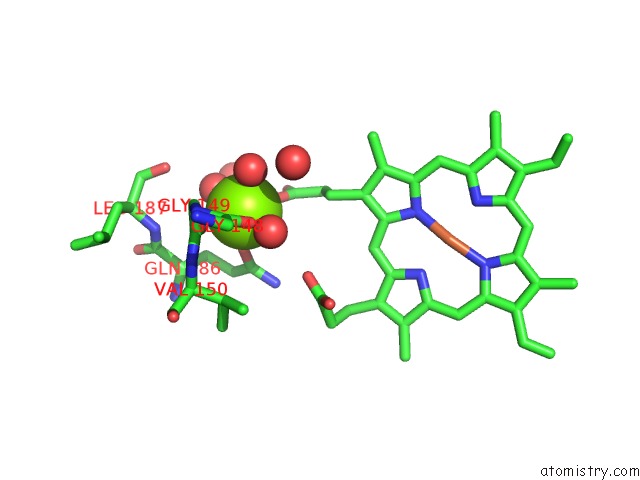
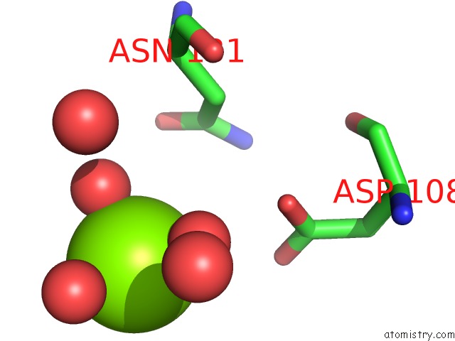
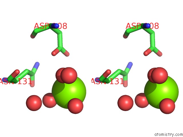
Magnesium binding site 1 out of 3 in 4qi3
Go back to
Magnesium binding site 1 out
of 3 in the Cytochrome Domain of Myriococcum Thermophilum Cellobiose Dehydrogenase, Mtcyt
Mono view
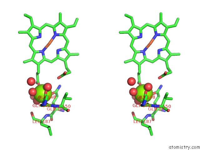
Stereo pair view
Mono view
Stereo pair view
A full contact list of Magnesium with other atoms in the Mg binding
site number 1 of Cytochrome Domain of Myriococcum Thermophilum Cellobiose Dehydrogenase, Mtcyt within 5.0Å range:
|
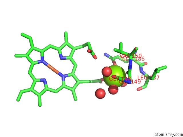
Magnesium binding site 2 out of 3 in 4qi3
Go back to
Magnesium binding site 2 out
of 3 in the Cytochrome Domain of Myriococcum Thermophilum Cellobiose Dehydrogenase, Mtcyt
Mono view
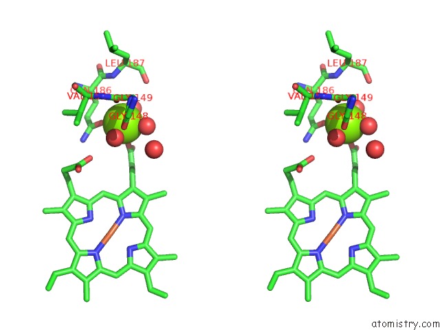
Stereo pair view
Mono view
Stereo pair view
A full contact list of Magnesium with other atoms in the Mg binding
site number 2 of Cytochrome Domain of Myriococcum Thermophilum Cellobiose Dehydrogenase, Mtcyt within 5.0Å range:
|
Magnesium binding site 3 out of 3 in 4qi3
Go back to
Magnesium binding site 3 out
of 3 in the Cytochrome Domain of Myriococcum Thermophilum Cellobiose Dehydrogenase, Mtcyt
Mono view
Stereo pair view
Mono view
Stereo pair view
A full contact list of Magnesium with other atoms in the Mg binding
site number 3 of Cytochrome Domain of Myriococcum Thermophilum Cellobiose Dehydrogenase, Mtcyt within 5.0Å range:
|
Reference:
T.C.Tan,
D.Kracher,
R.Gandini,
C.Sygmund,
R.Kittl,
D.Haltrich,
B.M.Hallberg,
R.Ludwig,
C.Divne.
Structural Basis For Cellobiose Dehydrogenase Action During Oxidative Cellulose Degradation. Nat Commun V. 6 7542 2015.
ISSN: ESSN 2041-1723
PubMed: 26151670
DOI: 10.1038/NCOMMS8542
Page generated: Tue Aug 20 01:59:40 2024
ISSN: ESSN 2041-1723
PubMed: 26151670
DOI: 10.1038/NCOMMS8542
Last articles
Fe in 5BYSFe in 5BYQ
Fe in 5BYR
Fe in 5C1V
Fe in 5C0Z
Fe in 5BVH
Fe in 5BVG
Fe in 5BWE
Fe in 5BWH
Fe in 5BWG