Magnesium »
PDB 4r17-4rcy »
4r4p »
Magnesium in PDB 4r4p: Crystal Structure of the Vs Ribozyme-A756G Mutant
Protein crystallography data
The structure of Crystal Structure of the Vs Ribozyme-A756G Mutant, PDB code: 4r4p
was solved by
J.A.Piccirilli,
N.B.Suslov,
S.Dasgupta,
H.Huang,
D.M.J.Lilley,
P.A.Rice,
with X-Ray Crystallography technique. A brief refinement statistics is given in the table below:
| Resolution Low / High (Å) | 41.47 / 3.07 |
| Space group | P 43 21 2 |
| Cell size a, b, c (Å), α, β, γ (°) | 102.680, 102.680, 211.021, 90.00, 90.00, 90.00 |
| R / Rfree (%) | 23.5 / 26.9 |
Magnesium Binding Sites:
The binding sites of Magnesium atom in the Crystal Structure of the Vs Ribozyme-A756G Mutant
(pdb code 4r4p). This binding sites where shown within
5.0 Angstroms radius around Magnesium atom.
In total 2 binding sites of Magnesium where determined in the Crystal Structure of the Vs Ribozyme-A756G Mutant, PDB code: 4r4p:
Jump to Magnesium binding site number: 1; 2;
In total 2 binding sites of Magnesium where determined in the Crystal Structure of the Vs Ribozyme-A756G Mutant, PDB code: 4r4p:
Jump to Magnesium binding site number: 1; 2;
Magnesium binding site 1 out of 2 in 4r4p
Go back to
Magnesium binding site 1 out
of 2 in the Crystal Structure of the Vs Ribozyme-A756G Mutant
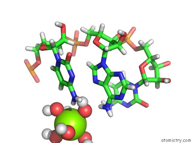
Mono view
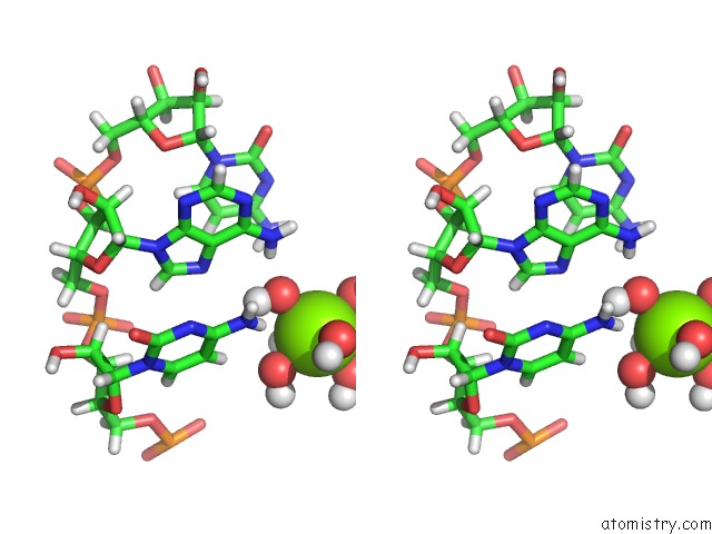
Stereo pair view
Mono view
Stereo pair view
A full contact list of Magnesium with other atoms in the Mg binding
site number 1 of Crystal Structure of the Vs Ribozyme-A756G Mutant within 5.0Å range:
|
Magnesium binding site 2 out of 2 in 4r4p
Go back to
Magnesium binding site 2 out
of 2 in the Crystal Structure of the Vs Ribozyme-A756G Mutant
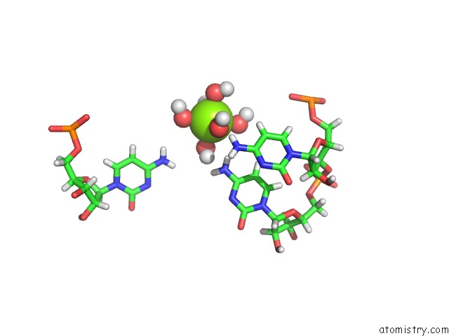
Mono view
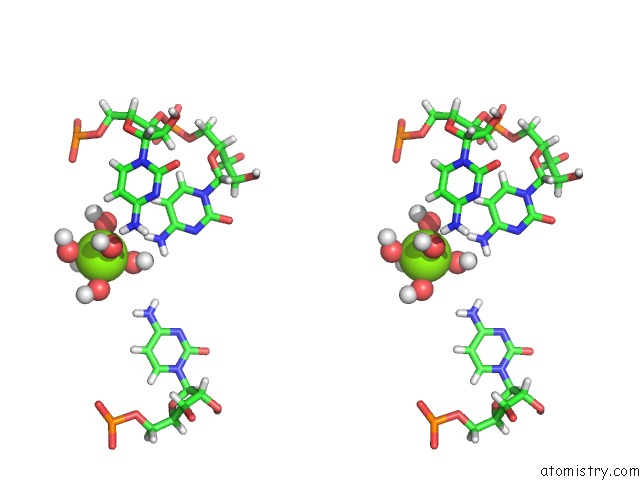
Stereo pair view
Mono view
Stereo pair view
A full contact list of Magnesium with other atoms in the Mg binding
site number 2 of Crystal Structure of the Vs Ribozyme-A756G Mutant within 5.0Å range:
|
Reference:
N.B.Suslov,
S.Dasgupta,
H.Huang,
J.R.Fuller,
D.M.Lilley,
P.A.Rice,
J.A.Piccirilli.
Crystal Structure of the Varkud Satellite Ribozyme. Nat.Chem.Biol. V. 11 840 2015.
ISSN: ISSN 1552-4450
PubMed: 26414446
DOI: 10.1038/NCHEMBIO.1929
Page generated: Mon Aug 11 23:08:52 2025
ISSN: ISSN 1552-4450
PubMed: 26414446
DOI: 10.1038/NCHEMBIO.1929
Last articles
Mg in 5G0RMg in 5G5V
Mg in 5G3T
Mg in 5G5T
Mg in 5G5S
Mg in 5G4A
Mg in 5G57
Mg in 5G50
Mg in 5G41
Mg in 5G3Z