Magnesium »
PDB 4r17-4rcy »
4r7t »
Magnesium in PDB 4r7t: Crystal Structure of Glucosamine-6-Phosphate Deaminase From Vibrio Cholerae
Enzymatic activity of Crystal Structure of Glucosamine-6-Phosphate Deaminase From Vibrio Cholerae
All present enzymatic activity of Crystal Structure of Glucosamine-6-Phosphate Deaminase From Vibrio Cholerae:
3.5.99.6;
3.5.99.6;
Protein crystallography data
The structure of Crystal Structure of Glucosamine-6-Phosphate Deaminase From Vibrio Cholerae, PDB code: 4r7t
was solved by
N.Maltseva,
Y.Kim,
K.Kwon,
W.F.Anderson,
A.Joachimiak,
Csgid,
Center Forstructural Genomics Of Infectious Diseases (Csgid),
with X-Ray Crystallography technique. A brief refinement statistics is given in the table below:
| Resolution Low / High (Å) | 38.65 / 2.10 |
| Space group | C 1 2 1 |
| Cell size a, b, c (Å), α, β, γ (°) | 78.359, 135.124, 86.001, 90.00, 106.15, 90.00 |
| R / Rfree (%) | 19.9 / 25.4 |
Other elements in 4r7t:
The structure of Crystal Structure of Glucosamine-6-Phosphate Deaminase From Vibrio Cholerae also contains other interesting chemical elements:
| Chlorine | (Cl) | 1 atom |
Magnesium Binding Sites:
The binding sites of Magnesium atom in the Crystal Structure of Glucosamine-6-Phosphate Deaminase From Vibrio Cholerae
(pdb code 4r7t). This binding sites where shown within
5.0 Angstroms radius around Magnesium atom.
In total 3 binding sites of Magnesium where determined in the Crystal Structure of Glucosamine-6-Phosphate Deaminase From Vibrio Cholerae, PDB code: 4r7t:
Jump to Magnesium binding site number: 1; 2; 3;
In total 3 binding sites of Magnesium where determined in the Crystal Structure of Glucosamine-6-Phosphate Deaminase From Vibrio Cholerae, PDB code: 4r7t:
Jump to Magnesium binding site number: 1; 2; 3;
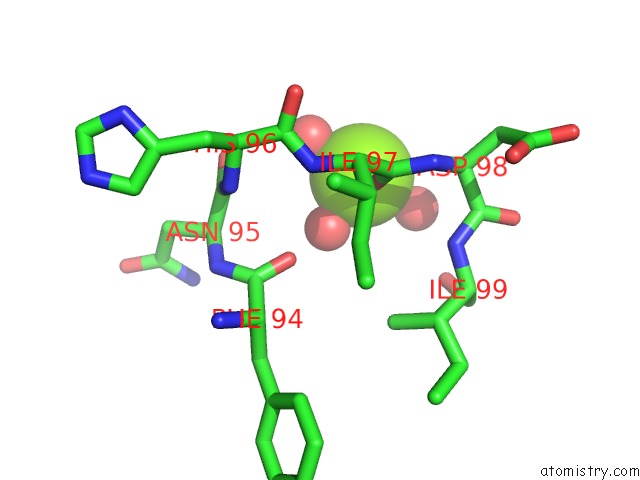
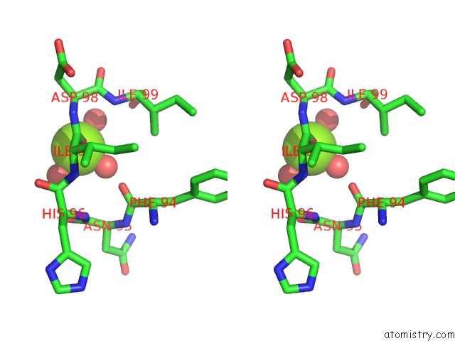
Magnesium binding site 1 out of 3 in 4r7t
Go back to
Magnesium binding site 1 out
of 3 in the Crystal Structure of Glucosamine-6-Phosphate Deaminase From Vibrio Cholerae
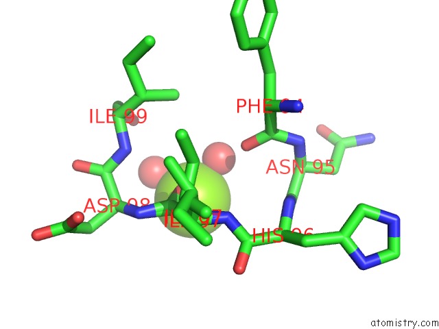
Mono view
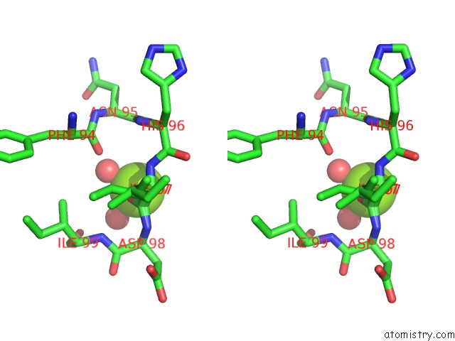
Stereo pair view
Mono view
Stereo pair view
A full contact list of Magnesium with other atoms in the Mg binding
site number 1 of Crystal Structure of Glucosamine-6-Phosphate Deaminase From Vibrio Cholerae within 5.0Å range:
|
Magnesium binding site 2 out of 3 in 4r7t
Go back to
Magnesium binding site 2 out
of 3 in the Crystal Structure of Glucosamine-6-Phosphate Deaminase From Vibrio Cholerae
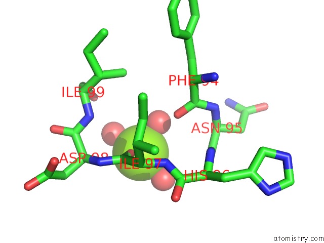
Mono view
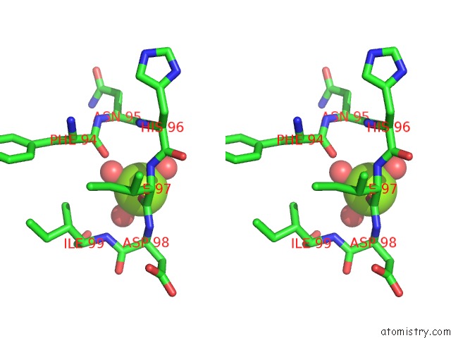
Stereo pair view
Mono view
Stereo pair view
A full contact list of Magnesium with other atoms in the Mg binding
site number 2 of Crystal Structure of Glucosamine-6-Phosphate Deaminase From Vibrio Cholerae within 5.0Å range:
|
Magnesium binding site 3 out of 3 in 4r7t
Go back to
Magnesium binding site 3 out
of 3 in the Crystal Structure of Glucosamine-6-Phosphate Deaminase From Vibrio Cholerae
Mono view
Stereo pair view
Mono view
Stereo pair view
A full contact list of Magnesium with other atoms in the Mg binding
site number 3 of Crystal Structure of Glucosamine-6-Phosphate Deaminase From Vibrio Cholerae within 5.0Å range:
|
Reference:
N.Maltseva,
Y.Kim,
K.Kwon,
W.F.Anderson,
A.Joachimiak,
Csgid.
Crystal Structure of Glucosamine-6-Phosphate Deaminase From Vibrio Cholerae To Be Published.
Page generated: Tue Aug 20 02:53:50 2024
Last articles
Mg in 3ONNMg in 3OMI
Mg in 3OMN
Mg in 3OMY
Mg in 3OL7
Mg in 3OMB
Mg in 3OMA
Mg in 3OM3
Mg in 3OM2
Mg in 3OLP