Magnesium »
PDB 4rrk-4s1l »
4rw3 »
Magnesium in PDB 4rw3: Structural Insights Into Substrate Binding of Brown Spider Venom Class II Phospholipases D
Enzymatic activity of Structural Insights Into Substrate Binding of Brown Spider Venom Class II Phospholipases D
All present enzymatic activity of Structural Insights Into Substrate Binding of Brown Spider Venom Class II Phospholipases D:
3.1.4.4;
3.1.4.4;
Protein crystallography data
The structure of Structural Insights Into Substrate Binding of Brown Spider Venom Class II Phospholipases D, PDB code: 4rw3
was solved by
M.A.Coronado,
A.Ullah,
L.S.Da Silva,
D.Chaves-Moreira,
L.Vuitika,
O.M.Chaim,
S.S.Veiga,
J.Chahine,
M.T.Murakami,
R.K.Arni,
with X-Ray Crystallography technique. A brief refinement statistics is given in the table below:
| Resolution Low / High (Å) | 30.00 / 1.72 |
| Space group | P 1 21 1 |
| Cell size a, b, c (Å), α, β, γ (°) | 49.810, 49.300, 56.300, 90.00, 105.83, 90.00 |
| R / Rfree (%) | 18.3 / 22.3 |
Magnesium Binding Sites:
The binding sites of Magnesium atom in the Structural Insights Into Substrate Binding of Brown Spider Venom Class II Phospholipases D
(pdb code 4rw3). This binding sites where shown within
5.0 Angstroms radius around Magnesium atom.
In total only one binding site of Magnesium was determined in the Structural Insights Into Substrate Binding of Brown Spider Venom Class II Phospholipases D, PDB code: 4rw3:
In total only one binding site of Magnesium was determined in the Structural Insights Into Substrate Binding of Brown Spider Venom Class II Phospholipases D, PDB code: 4rw3:
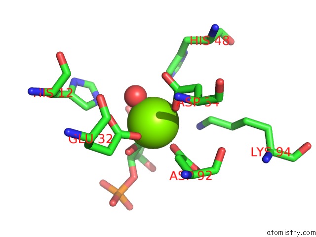
Magnesium binding site 1 out of 1 in 4rw3
Go back to
Magnesium binding site 1 out
of 1 in the Structural Insights Into Substrate Binding of Brown Spider Venom Class II Phospholipases D
Mono view
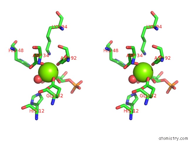
Stereo pair view
Mono view
Stereo pair view
A full contact list of Magnesium with other atoms in the Mg binding
site number 1 of Structural Insights Into Substrate Binding of Brown Spider Venom Class II Phospholipases D within 5.0Å range:
|
Reference:
M.A.Coronado,
A.Ullah,
L.S.Da Silva,
D.Chaves-Moreira,
L.Vuitika,
O.M.Chaim,
S.S.Veiga,
J.Chahine,
M.T.Murakami,
R.K.Arni.
Structural Insights Into Substrate Binding of Brown Spider Venom Class II Phospholipases D. Curr Protein Pept Sci V. 16 768 2015.
PubMed: 25961401
Page generated: Tue Aug 20 03:31:56 2024
PubMed: 25961401
Last articles
Mg in 3BC1Mg in 3BB1
Mg in 3BBF
Mg in 3B9T
Mg in 3BBP
Mg in 3B97
Mg in 3BB4
Mg in 3BB3
Mg in 3B9R
Mg in 3B8I