Magnesium »
PDB 4rrk-4s1l »
4rwt »
Magnesium in PDB 4rwt: Structure of Actin-Lmod Complex
Protein crystallography data
The structure of Structure of Actin-Lmod Complex, PDB code: 4rwt
was solved by
X.Chen,
F.Ni,
Q.Wang,
with X-Ray Crystallography technique. A brief refinement statistics is given in the table below:
| Resolution Low / High (Å) | 51.07 / 2.98 |
| Space group | P 1 |
| Cell size a, b, c (Å), α, β, γ (°) | 65.350, 65.650, 81.920, 101.29, 90.94, 107.97 |
| R / Rfree (%) | 24.8 / 25.7 |
Magnesium Binding Sites:
The binding sites of Magnesium atom in the Structure of Actin-Lmod Complex
(pdb code 4rwt). This binding sites where shown within
5.0 Angstroms radius around Magnesium atom.
In total 2 binding sites of Magnesium where determined in the Structure of Actin-Lmod Complex, PDB code: 4rwt:
Jump to Magnesium binding site number: 1; 2;
In total 2 binding sites of Magnesium where determined in the Structure of Actin-Lmod Complex, PDB code: 4rwt:
Jump to Magnesium binding site number: 1; 2;
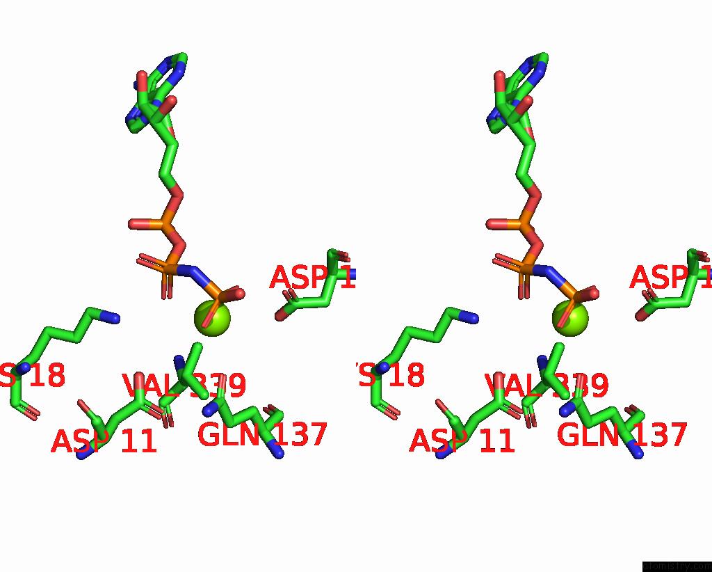
Magnesium binding site 1 out of 2 in 4rwt
Go back to
Magnesium binding site 1 out
of 2 in the Structure of Actin-Lmod Complex

Mono view

Stereo pair view
Mono view
Stereo pair view
A full contact list of Magnesium with other atoms in the Mg binding
site number 1 of Structure of Actin-Lmod Complex within 5.0Å range:
|
Magnesium binding site 2 out of 2 in 4rwt
Go back to
Magnesium binding site 2 out
of 2 in the Structure of Actin-Lmod Complex

Mono view
Stereo pair view
Mono view
Stereo pair view
A full contact list of Magnesium with other atoms in the Mg binding
site number 2 of Structure of Actin-Lmod Complex within 5.0Å range:
|
Reference:
X.Chen,
F.Ni,
E.Kondrashkina,
J.Ma,
Q.Wang.
Mechanisms of Leiomodin 2-Mediated Regulation of Actin Filament in Muscle Cells. Proc.Natl.Acad.Sci.Usa V. 112 12687 2015.
ISSN: ISSN 0027-8424
PubMed: 26417072
DOI: 10.1073/PNAS.1512464112
Page generated: Tue Aug 20 03:32:54 2024
ISSN: ISSN 0027-8424
PubMed: 26417072
DOI: 10.1073/PNAS.1512464112
Last articles
Mg in 3BC1Mg in 3BB1
Mg in 3BBF
Mg in 3B9T
Mg in 3BBP
Mg in 3B97
Mg in 3BB4
Mg in 3BB3
Mg in 3B9R
Mg in 3B8I