Magnesium »
PDB 4rrk-4s1l »
4s1k »
Magnesium in PDB 4s1k: Structure of Uranotaenia Sapphirina Cypovirus (CPV17) Polyhedrin at 100 K
Protein crystallography data
The structure of Structure of Uranotaenia Sapphirina Cypovirus (CPV17) Polyhedrin at 100 K, PDB code: 4s1k
was solved by
H.M.Ginn,
M.Messerschmidt,
X.Ji,
H.Zhang,
D.Axford,
R.J.Gildea,
G.Winter,
A.S.Brewster,
J.Hattne,
A.Wagner,
J.M.Grimes,
G.Evans,
N.K.Sauter,
G.Sutton,
D.I.Stuart,
with X-Ray Crystallography technique. A brief refinement statistics is given in the table below:
| Resolution Low / High (Å) | 74.00 / 2.20 |
| Space group | I 2 3 |
| Cell size a, b, c (Å), α, β, γ (°) | 104.879, 104.879, 104.879, 90.00, 90.00, 90.00 |
| R / Rfree (%) | 14.8 / 19.9 |
Magnesium Binding Sites:
The binding sites of Magnesium atom in the Structure of Uranotaenia Sapphirina Cypovirus (CPV17) Polyhedrin at 100 K
(pdb code 4s1k). This binding sites where shown within
5.0 Angstroms radius around Magnesium atom.
In total only one binding site of Magnesium was determined in the Structure of Uranotaenia Sapphirina Cypovirus (CPV17) Polyhedrin at 100 K, PDB code: 4s1k:
In total only one binding site of Magnesium was determined in the Structure of Uranotaenia Sapphirina Cypovirus (CPV17) Polyhedrin at 100 K, PDB code: 4s1k:
Magnesium binding site 1 out of 1 in 4s1k
Go back to
Magnesium binding site 1 out
of 1 in the Structure of Uranotaenia Sapphirina Cypovirus (CPV17) Polyhedrin at 100 K
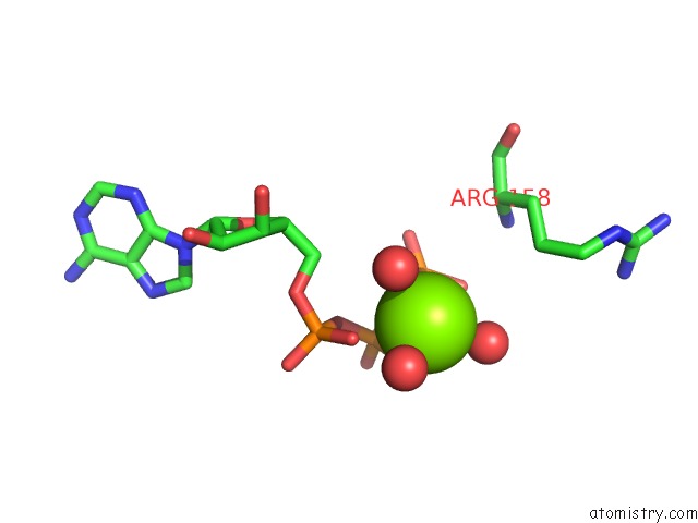
Mono view
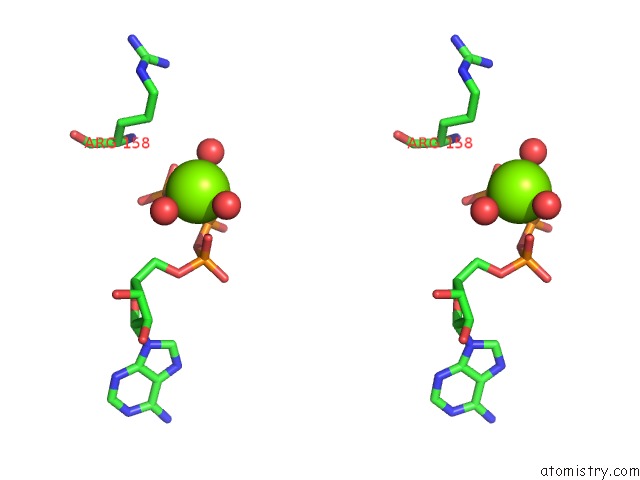
Stereo pair view
Mono view
Stereo pair view
A full contact list of Magnesium with other atoms in the Mg binding
site number 1 of Structure of Uranotaenia Sapphirina Cypovirus (CPV17) Polyhedrin at 100 K within 5.0Å range:
|
Reference:
H.M.Ginn,
M.Messerschmidt,
X.Ji,
H.Zhang,
D.Axford,
R.J.Gildea,
G.Winter,
A.S.Brewster,
J.Hattne,
A.Wagner,
J.M.Grimes,
G.Evans,
N.K.Sauter,
G.Sutton,
D.I.Stuart.
Structure of CPV17 Polyhedrin Determined By the Improved Analysis of Serial Femtosecond Crystallographic Data. Nat Commun V. 6 6435 2015.
ISSN: ESSN 2041-1723
PubMed: 25751308
DOI: 10.1038/NCOMMS7435
Page generated: Tue Aug 20 03:38:45 2024
ISSN: ESSN 2041-1723
PubMed: 25751308
DOI: 10.1038/NCOMMS7435
Last articles
Mg in 4K6OMg in 4K9P
Mg in 4K9O
Mg in 4K9M
Mg in 4K9K
Mg in 4K99
Mg in 4K98
Mg in 4K97
Mg in 4K8Z
Mg in 4K8O