Magnesium »
PDB 4u9i-4um5 »
4uau »
Magnesium in PDB 4uau: Crystal Structure of Cbby (Mutant D10N) From Rhodobacter Sphaeroides in Complex with Xylulose-(1,5)Bisphosphate, Crystal Form II
Protein crystallography data
The structure of Crystal Structure of Cbby (Mutant D10N) From Rhodobacter Sphaeroides in Complex with Xylulose-(1,5)Bisphosphate, Crystal Form II, PDB code: 4uau
was solved by
A.Bracher,
A.Sharma,
A.Starling-Windhof,
F.U.Hartl,
M.Hayer-Hartl,
with X-Ray Crystallography technique. A brief refinement statistics is given in the table below:
| Resolution Low / High (Å) | 30.00 / 1.45 |
| Space group | P 21 21 21 |
| Cell size a, b, c (Å), α, β, γ (°) | 51.288, 69.767, 125.793, 90.00, 90.00, 90.00 |
| R / Rfree (%) | 17.9 / 21.1 |
Magnesium Binding Sites:
The binding sites of Magnesium atom in the Crystal Structure of Cbby (Mutant D10N) From Rhodobacter Sphaeroides in Complex with Xylulose-(1,5)Bisphosphate, Crystal Form II
(pdb code 4uau). This binding sites where shown within
5.0 Angstroms radius around Magnesium atom.
In total 2 binding sites of Magnesium where determined in the Crystal Structure of Cbby (Mutant D10N) From Rhodobacter Sphaeroides in Complex with Xylulose-(1,5)Bisphosphate, Crystal Form II, PDB code: 4uau:
Jump to Magnesium binding site number: 1; 2;
In total 2 binding sites of Magnesium where determined in the Crystal Structure of Cbby (Mutant D10N) From Rhodobacter Sphaeroides in Complex with Xylulose-(1,5)Bisphosphate, Crystal Form II, PDB code: 4uau:
Jump to Magnesium binding site number: 1; 2;
Magnesium binding site 1 out of 2 in 4uau
Go back to
Magnesium binding site 1 out
of 2 in the Crystal Structure of Cbby (Mutant D10N) From Rhodobacter Sphaeroides in Complex with Xylulose-(1,5)Bisphosphate, Crystal Form II

Mono view
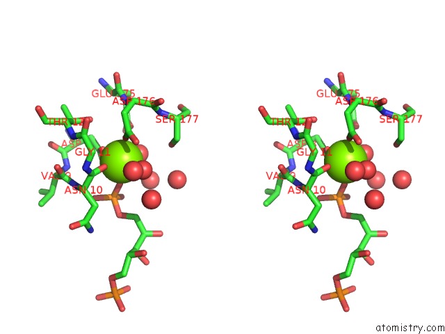
Stereo pair view
Mono view
Stereo pair view
A full contact list of Magnesium with other atoms in the Mg binding
site number 1 of Crystal Structure of Cbby (Mutant D10N) From Rhodobacter Sphaeroides in Complex with Xylulose-(1,5)Bisphosphate, Crystal Form II within 5.0Å range:
|
Magnesium binding site 2 out of 2 in 4uau
Go back to
Magnesium binding site 2 out
of 2 in the Crystal Structure of Cbby (Mutant D10N) From Rhodobacter Sphaeroides in Complex with Xylulose-(1,5)Bisphosphate, Crystal Form II
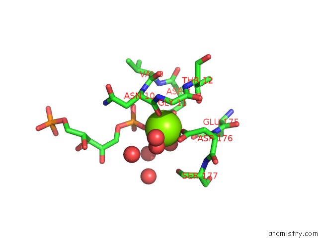
Mono view

Stereo pair view
Mono view
Stereo pair view
A full contact list of Magnesium with other atoms in the Mg binding
site number 2 of Crystal Structure of Cbby (Mutant D10N) From Rhodobacter Sphaeroides in Complex with Xylulose-(1,5)Bisphosphate, Crystal Form II within 5.0Å range:
|
Reference:
A.Bracher,
A.Sharma,
A.Starling-Windhof,
F.U.Hartl,
M.Hayer-Hartl.
Degradation of Potent Rubisco Inhibitor By Selective Sugar Phosphatase Nat.Plants 2015.
DOI: 10.1038/NPLANTS.2014.2
Page generated: Tue Aug 20 04:34:12 2024
DOI: 10.1038/NPLANTS.2014.2
Last articles
K in 8HK6K in 8HKK
K in 8HKF
K in 8HKM
K in 8HIR
K in 8GYR
K in 8GR9
K in 8H5L
K in 8H5J
K in 8GPE