Magnesium »
PDB 4u9i-4um5 »
4ueg »
Magnesium in PDB 4ueg: Crystal Structure of Human Glycogenin-2 Catalytic Domain
Enzymatic activity of Crystal Structure of Human Glycogenin-2 Catalytic Domain
All present enzymatic activity of Crystal Structure of Human Glycogenin-2 Catalytic Domain:
2.4.1.186;
2.4.1.186;
Protein crystallography data
The structure of Crystal Structure of Human Glycogenin-2 Catalytic Domain, PDB code: 4ueg
was solved by
M.Fairhead,
C.Strain-Damerell,
T.Krojer,
D.S.Froese,
J.Kopec,
R.Nowak,
N.Burgess-Brown,
F.Von Delft,
C.Arrowsmith,
A.Edwards,
C.Bountra,
W.W.Yue,
with X-Ray Crystallography technique. A brief refinement statistics is given in the table below:
| Resolution Low / High (Å) | 64.608 / 1.93 |
| Space group | C 1 2 1 |
| Cell size a, b, c (Å), α, β, γ (°) | 127.113, 80.541, 73.683, 90.00, 118.74, 90.00 |
| R / Rfree (%) | 18.67 / 22.43 |
Magnesium Binding Sites:
The binding sites of Magnesium atom in the Crystal Structure of Human Glycogenin-2 Catalytic Domain
(pdb code 4ueg). This binding sites where shown within
5.0 Angstroms radius around Magnesium atom.
In total 2 binding sites of Magnesium where determined in the Crystal Structure of Human Glycogenin-2 Catalytic Domain, PDB code: 4ueg:
Jump to Magnesium binding site number: 1; 2;
In total 2 binding sites of Magnesium where determined in the Crystal Structure of Human Glycogenin-2 Catalytic Domain, PDB code: 4ueg:
Jump to Magnesium binding site number: 1; 2;
Magnesium binding site 1 out of 2 in 4ueg
Go back to
Magnesium binding site 1 out
of 2 in the Crystal Structure of Human Glycogenin-2 Catalytic Domain
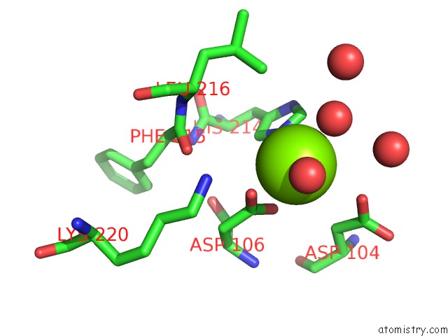
Mono view
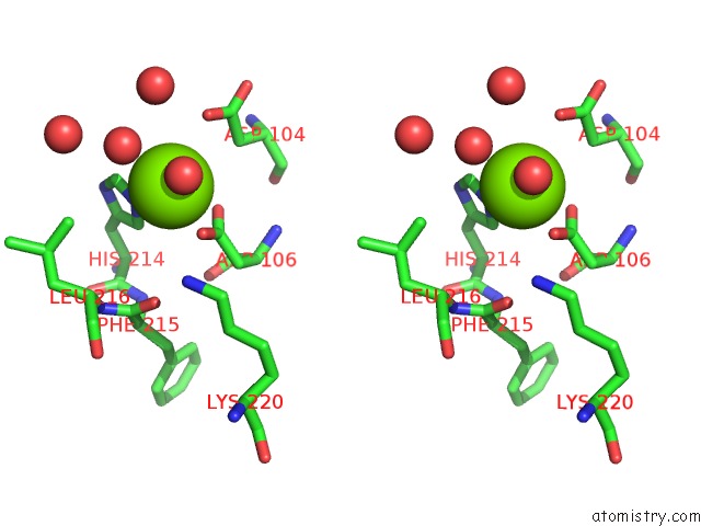
Stereo pair view
Mono view
Stereo pair view
A full contact list of Magnesium with other atoms in the Mg binding
site number 1 of Crystal Structure of Human Glycogenin-2 Catalytic Domain within 5.0Å range:
|
Magnesium binding site 2 out of 2 in 4ueg
Go back to
Magnesium binding site 2 out
of 2 in the Crystal Structure of Human Glycogenin-2 Catalytic Domain
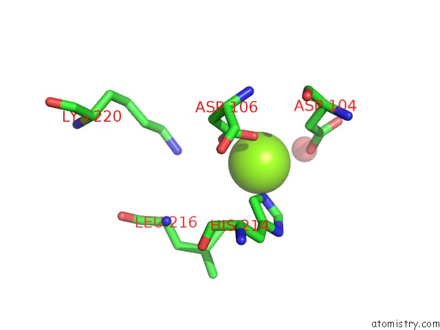
Mono view
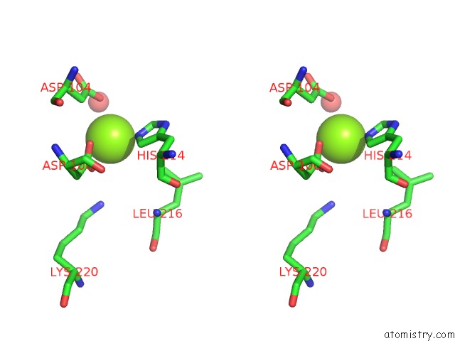
Stereo pair view
Mono view
Stereo pair view
A full contact list of Magnesium with other atoms in the Mg binding
site number 2 of Crystal Structure of Human Glycogenin-2 Catalytic Domain within 5.0Å range:
|
Reference:
M.Fairhead,
C.Strain-Damerell,
T.Krojer,
D.S.Froese,
J.Kopec,
R.Nowak,
N.Burgess-Brown,
F.Von Delft,
C.Arrowsmith,
A.Edwards,
C.Bountra,
W.W.Yue.
Crystal Structure of Human Glycogenin-2 Catalytic Domain To Be Published.
Page generated: Tue Aug 20 04:40:05 2024
Last articles
K in 7OA3K in 7OAR
K in 7O5R
K in 7O6R
K in 7OA2
K in 7NNL
K in 7NY1
K in 7O2P
K in 7O5I
K in 7O2R