Magnesium »
PDB 5ac1-5avy »
5aes »
Magnesium in PDB 5aes: Crystal Structure of Murine Chronophin (Pyridoxal Phosphate Phosphatase) in Complex with A Pnp-Derived Inhibitor
Enzymatic activity of Crystal Structure of Murine Chronophin (Pyridoxal Phosphate Phosphatase) in Complex with A Pnp-Derived Inhibitor
All present enzymatic activity of Crystal Structure of Murine Chronophin (Pyridoxal Phosphate Phosphatase) in Complex with A Pnp-Derived Inhibitor:
3.1.3.74;
3.1.3.74;
Protein crystallography data
The structure of Crystal Structure of Murine Chronophin (Pyridoxal Phosphate Phosphatase) in Complex with A Pnp-Derived Inhibitor, PDB code: 5aes
was solved by
G.Knobloch,
N.Jabari,
M.Koehn,
A.Gohla,
H.Schindelin,
with X-Ray Crystallography technique. A brief refinement statistics is given in the table below:
| Resolution Low / High (Å) | 32.714 / 2.75 |
| Space group | I 2 3 |
| Cell size a, b, c (Å), α, β, γ (°) | 166.810, 166.810, 166.810, 90.00, 90.00, 90.00 |
| R / Rfree (%) | 18.95 / 24.66 |
Magnesium Binding Sites:
The binding sites of Magnesium atom in the Crystal Structure of Murine Chronophin (Pyridoxal Phosphate Phosphatase) in Complex with A Pnp-Derived Inhibitor
(pdb code 5aes). This binding sites where shown within
5.0 Angstroms radius around Magnesium atom.
In total 2 binding sites of Magnesium where determined in the Crystal Structure of Murine Chronophin (Pyridoxal Phosphate Phosphatase) in Complex with A Pnp-Derived Inhibitor, PDB code: 5aes:
Jump to Magnesium binding site number: 1; 2;
In total 2 binding sites of Magnesium where determined in the Crystal Structure of Murine Chronophin (Pyridoxal Phosphate Phosphatase) in Complex with A Pnp-Derived Inhibitor, PDB code: 5aes:
Jump to Magnesium binding site number: 1; 2;
Magnesium binding site 1 out of 2 in 5aes
Go back to
Magnesium binding site 1 out
of 2 in the Crystal Structure of Murine Chronophin (Pyridoxal Phosphate Phosphatase) in Complex with A Pnp-Derived Inhibitor
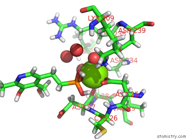
Mono view
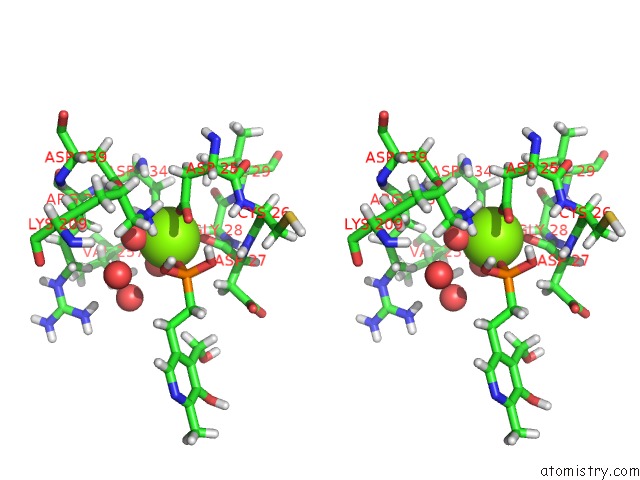
Stereo pair view
Mono view
Stereo pair view
A full contact list of Magnesium with other atoms in the Mg binding
site number 1 of Crystal Structure of Murine Chronophin (Pyridoxal Phosphate Phosphatase) in Complex with A Pnp-Derived Inhibitor within 5.0Å range:
|
Magnesium binding site 2 out of 2 in 5aes
Go back to
Magnesium binding site 2 out
of 2 in the Crystal Structure of Murine Chronophin (Pyridoxal Phosphate Phosphatase) in Complex with A Pnp-Derived Inhibitor
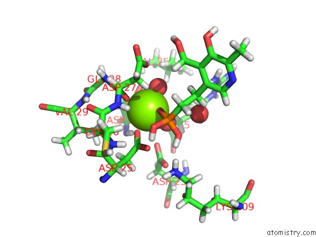
Mono view
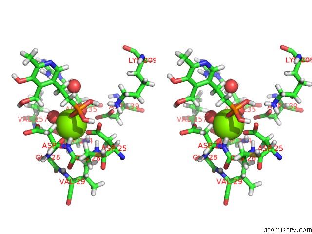
Stereo pair view
Mono view
Stereo pair view
A full contact list of Magnesium with other atoms in the Mg binding
site number 2 of Crystal Structure of Murine Chronophin (Pyridoxal Phosphate Phosphatase) in Complex with A Pnp-Derived Inhibitor within 5.0Å range:
|
Reference:
G.Knobloch,
N.Jabari,
S.Stadlbauer,
H.Schindelin,
M.Kohn,
A.Gohla.
Synthesis of Hydrolysis-Resistant Pyridoxal 5'-Phosphate Analogs and Their Biochemical and X-Ray Crystallographic Characterization with the Pyridoxal Phosphatase Chronophin. Bioorg.Med.Chem. 2015.
ISSN: ESSN 1464-3391
PubMed: 25783190
DOI: 10.1016/J.BMC.2015.02.049
Page generated: Tue Aug 12 05:09:34 2025
ISSN: ESSN 1464-3391
PubMed: 25783190
DOI: 10.1016/J.BMC.2015.02.049
Last articles
Mg in 5GG8Mg in 5GG7
Mg in 5GG6
Mg in 5GAD
Mg in 5GAG
Mg in 5GAF
Mg in 5GAE
Mg in 5G0R
Mg in 5G5V
Mg in 5G3T