Magnesium »
PDB 5ca3-5chg »
5cak »
Magnesium in PDB 5cak: Crystal Structure of Mycobacterium Tuberculosis Malate Synthase in Complex with 2-Hydroxy-3-(1H-Indol-3-Yl)Propanoic Acid
Enzymatic activity of Crystal Structure of Mycobacterium Tuberculosis Malate Synthase in Complex with 2-Hydroxy-3-(1H-Indol-3-Yl)Propanoic Acid
All present enzymatic activity of Crystal Structure of Mycobacterium Tuberculosis Malate Synthase in Complex with 2-Hydroxy-3-(1H-Indol-3-Yl)Propanoic Acid:
2.3.3.9;
2.3.3.9;
Protein crystallography data
The structure of Crystal Structure of Mycobacterium Tuberculosis Malate Synthase in Complex with 2-Hydroxy-3-(1H-Indol-3-Yl)Propanoic Acid, PDB code: 5cak
was solved by
H.-L.Huang,
J.C.Sacchettini,
with X-Ray Crystallography technique. A brief refinement statistics is given in the table below:
| Resolution Low / High (Å) | 49.58 / 1.99 |
| Space group | P 43 21 2 |
| Cell size a, b, c (Å), α, β, γ (°) | 78.238, 78.238, 223.566, 90.00, 90.00, 90.00 |
| R / Rfree (%) | 16.4 / 22.1 |
Magnesium Binding Sites:
The binding sites of Magnesium atom in the Crystal Structure of Mycobacterium Tuberculosis Malate Synthase in Complex with 2-Hydroxy-3-(1H-Indol-3-Yl)Propanoic Acid
(pdb code 5cak). This binding sites where shown within
5.0 Angstroms radius around Magnesium atom.
In total only one binding site of Magnesium was determined in the Crystal Structure of Mycobacterium Tuberculosis Malate Synthase in Complex with 2-Hydroxy-3-(1H-Indol-3-Yl)Propanoic Acid, PDB code: 5cak:
In total only one binding site of Magnesium was determined in the Crystal Structure of Mycobacterium Tuberculosis Malate Synthase in Complex with 2-Hydroxy-3-(1H-Indol-3-Yl)Propanoic Acid, PDB code: 5cak:
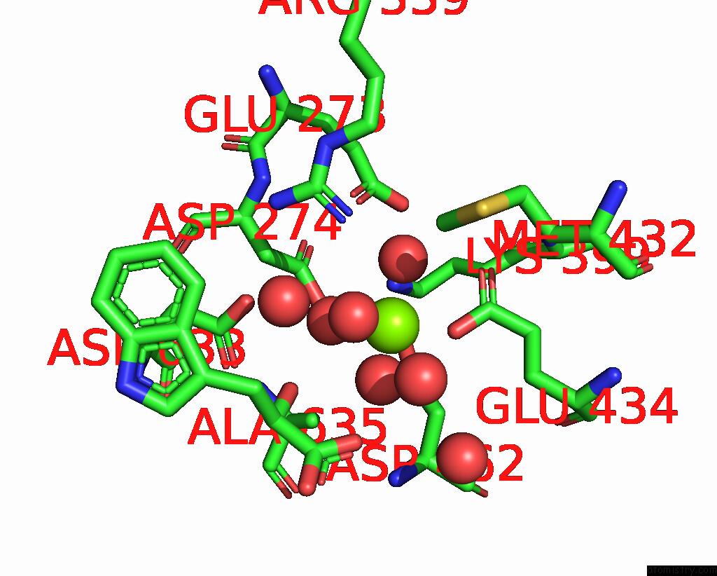
Magnesium binding site 1 out of 1 in 5cak
Go back to
Magnesium binding site 1 out
of 1 in the Crystal Structure of Mycobacterium Tuberculosis Malate Synthase in Complex with 2-Hydroxy-3-(1H-Indol-3-Yl)Propanoic Acid
Mono view

Stereo pair view
Mono view
Stereo pair view
A full contact list of Magnesium with other atoms in the Mg binding
site number 1 of Crystal Structure of Mycobacterium Tuberculosis Malate Synthase in Complex with 2-Hydroxy-3-(1H-Indol-3-Yl)Propanoic Acid within 5.0Å range:
|
Reference:
H.L.Huang,
I.V.Krieger,
M.K.Parai,
V.B.Gawandi,
J.C.Sacchettini.
Mycobacterium Tuberculosis Malate Synthase Structures with Fragments Reveal A Portal For Substrate/Product Exchange. J. Biol. Chem. V. 291 27421 2016.
ISSN: ESSN 1083-351X
PubMed: 27738104
DOI: 10.1074/JBC.M116.750877
Page generated: Sun Sep 29 02:02:14 2024
ISSN: ESSN 1083-351X
PubMed: 27738104
DOI: 10.1074/JBC.M116.750877
Last articles
Mg in 4DV5Mg in 4DV4
Mg in 4DV3
Mg in 4DV2
Mg in 4DV0
Mg in 4DV1
Mg in 4DUZ
Mg in 4DUY
Mg in 4DR7
Mg in 4DR6