Magnesium »
PDB 5ca3-5chg »
5cfg »
Magnesium in PDB 5cfg: C2 Crystal Form of APE1 with MG2+
Enzymatic activity of C2 Crystal Form of APE1 with MG2+
All present enzymatic activity of C2 Crystal Form of APE1 with MG2+:
4.2.99.18;
4.2.99.18;
Protein crystallography data
The structure of C2 Crystal Form of APE1 with MG2+, PDB code: 5cfg
was solved by
S.Morera,
A.Vigouroux,
with X-Ray Crystallography technique. A brief refinement statistics is given in the table below:
| Resolution Low / High (Å) | 41.39 / 1.80 |
| Space group | C 1 2 1 |
| Cell size a, b, c (Å), α, β, γ (°) | 86.530, 45.120, 77.980, 90.00, 105.15, 90.00 |
| R / Rfree (%) | 18.5 / 20.4 |
Magnesium Binding Sites:
The binding sites of Magnesium atom in the C2 Crystal Form of APE1 with MG2+
(pdb code 5cfg). This binding sites where shown within
5.0 Angstroms radius around Magnesium atom.
In total only one binding site of Magnesium was determined in the C2 Crystal Form of APE1 with MG2+, PDB code: 5cfg:
In total only one binding site of Magnesium was determined in the C2 Crystal Form of APE1 with MG2+, PDB code: 5cfg:
Magnesium binding site 1 out of 1 in 5cfg
Go back to
Magnesium binding site 1 out
of 1 in the C2 Crystal Form of APE1 with MG2+
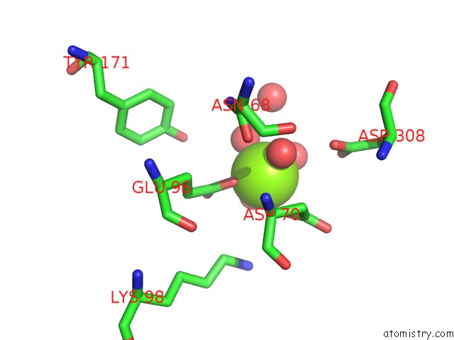
Mono view
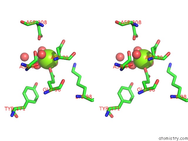
Stereo pair view
Mono view
Stereo pair view
A full contact list of Magnesium with other atoms in the Mg binding
site number 1 of C2 Crystal Form of APE1 with MG2+ within 5.0Å range:
|
Reference:
M.Redrejo-Rodriguez,
A.Vigouroux,
A.Mursalimov,
I.Grin,
D.Alili,
Z.Koshenov,
Z.Akishev,
A.Maksimenko,
A.K.Bissenbaev,
B.T.Matkarimov,
M.Saparbaev,
A.A.Ishchenko,
S.Morera.
Structural Comparison of Ap Endonucleases From the Exonuclease III Family Reveals New Amino Acid Residues in Human Ap Endonuclease 1 That Are Involved in Incision of Damaged Dna. Biochimie V.-129 20 2016.
ISSN: ISSN 0300-9084
PubMed: 27343627
DOI: 10.1016/J.BIOCHI.2016.06.011
Page generated: Sun Sep 29 02:04:53 2024
ISSN: ISSN 0300-9084
PubMed: 27343627
DOI: 10.1016/J.BIOCHI.2016.06.011
Last articles
Mg in 4DV5Mg in 4DV4
Mg in 4DV3
Mg in 4DV2
Mg in 4DV0
Mg in 4DV1
Mg in 4DUZ
Mg in 4DUY
Mg in 4DR7
Mg in 4DR6