Magnesium »
PDB 5cr7-5d2j »
5cx7 »
Magnesium in PDB 5cx7: Crystal Structure of Pduoc:Heme Complex
Protein crystallography data
The structure of Crystal Structure of Pduoc:Heme Complex, PDB code: 5cx7
was solved by
S.Geremia,
N.Hickey,
D.Ortiz De Orue Lucana,
with X-Ray Crystallography technique. A brief refinement statistics is given in the table below:
| Resolution Low / High (Å) | 33.07 / 1.97 |
| Space group | P 1 21 1 |
| Cell size a, b, c (Å), α, β, γ (°) | 71.300, 130.120, 120.750, 90.00, 90.00, 90.00 |
| R / Rfree (%) | 15.8 / 18.6 |
Other elements in 5cx7:
The structure of Crystal Structure of Pduoc:Heme Complex also contains other interesting chemical elements:
| Iron | (Fe) | 8 atoms |
| Chlorine | (Cl) | 4 atoms |
| Sodium | (Na) | 18 atoms |
Magnesium Binding Sites:
The binding sites of Magnesium atom in the Crystal Structure of Pduoc:Heme Complex
(pdb code 5cx7). This binding sites where shown within
5.0 Angstroms radius around Magnesium atom.
In total 2 binding sites of Magnesium where determined in the Crystal Structure of Pduoc:Heme Complex, PDB code: 5cx7:
Jump to Magnesium binding site number: 1; 2;
In total 2 binding sites of Magnesium where determined in the Crystal Structure of Pduoc:Heme Complex, PDB code: 5cx7:
Jump to Magnesium binding site number: 1; 2;
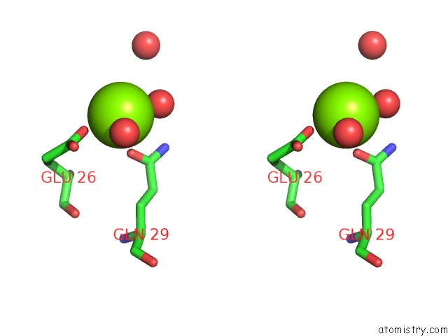
Magnesium binding site 1 out of 2 in 5cx7
Go back to
Magnesium binding site 1 out
of 2 in the Crystal Structure of Pduoc:Heme Complex

Mono view
Stereo pair view
Mono view
Stereo pair view
A full contact list of Magnesium with other atoms in the Mg binding
site number 1 of Crystal Structure of Pduoc:Heme Complex within 5.0Å range:
|
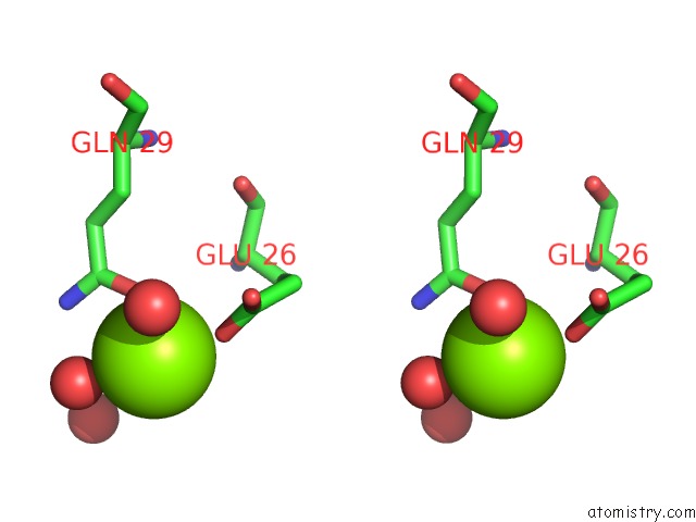
Magnesium binding site 2 out of 2 in 5cx7
Go back to
Magnesium binding site 2 out
of 2 in the Crystal Structure of Pduoc:Heme Complex

Mono view
Stereo pair view
Mono view
Stereo pair view
A full contact list of Magnesium with other atoms in the Mg binding
site number 2 of Crystal Structure of Pduoc:Heme Complex within 5.0Å range:
|
Reference:
D.Ortiz De Orue Lucana,
N.Hickey,
M.Hensel,
J.P.Klare,
S.Geremia,
T.Tiufiakova,
A.E.Torda.
The Crystal Structure of the C-Terminal Domain of the Salmonella Enterica Pduo Protein: An Old Fold with A New Heme-Binding Mode. Front Microbiol V. 7 1010 2016.
ISSN: ESSN 1664-302X
PubMed: 27446048
DOI: 10.3389/FMICB.2016.01010
Page generated: Sun Sep 29 02:19:19 2024
ISSN: ESSN 1664-302X
PubMed: 27446048
DOI: 10.3389/FMICB.2016.01010
Last articles
K in 9DITK in 9DJV
K in 9DIG
K in 9DID
K in 9DIC
K in 9DI8
K in 9DIB
K in 9DE5
K in 9D5W
K in 9D19