Magnesium »
PDB 5dt3-5e79 »
5dxi »
Magnesium in PDB 5dxi: Structure of C. Albicans Trehalose-6-Phosphate Phosphatase C-Terminal Domain
Enzymatic activity of Structure of C. Albicans Trehalose-6-Phosphate Phosphatase C-Terminal Domain
All present enzymatic activity of Structure of C. Albicans Trehalose-6-Phosphate Phosphatase C-Terminal Domain:
2.4.1.15;
2.4.1.15;
Protein crystallography data
The structure of Structure of C. Albicans Trehalose-6-Phosphate Phosphatase C-Terminal Domain, PDB code: 5dxi
was solved by
Y.Miao,
R.G.Brennan,
with X-Ray Crystallography technique. A brief refinement statistics is given in the table below:
| Resolution Low / High (Å) | 36.43 / 2.00 |
| Space group | P 21 21 21 |
| Cell size a, b, c (Å), α, β, γ (°) | 58.757, 87.200, 139.271, 90.00, 90.00, 90.00 |
| R / Rfree (%) | 20 / 24.2 |
Other elements in 5dxi:
The structure of Structure of C. Albicans Trehalose-6-Phosphate Phosphatase C-Terminal Domain also contains other interesting chemical elements:
| Fluorine | (F) | 6 atoms |
Magnesium Binding Sites:
The binding sites of Magnesium atom in the Structure of C. Albicans Trehalose-6-Phosphate Phosphatase C-Terminal Domain
(pdb code 5dxi). This binding sites where shown within
5.0 Angstroms radius around Magnesium atom.
In total 2 binding sites of Magnesium where determined in the Structure of C. Albicans Trehalose-6-Phosphate Phosphatase C-Terminal Domain, PDB code: 5dxi:
Jump to Magnesium binding site number: 1; 2;
In total 2 binding sites of Magnesium where determined in the Structure of C. Albicans Trehalose-6-Phosphate Phosphatase C-Terminal Domain, PDB code: 5dxi:
Jump to Magnesium binding site number: 1; 2;
Magnesium binding site 1 out of 2 in 5dxi
Go back to
Magnesium binding site 1 out
of 2 in the Structure of C. Albicans Trehalose-6-Phosphate Phosphatase C-Terminal Domain
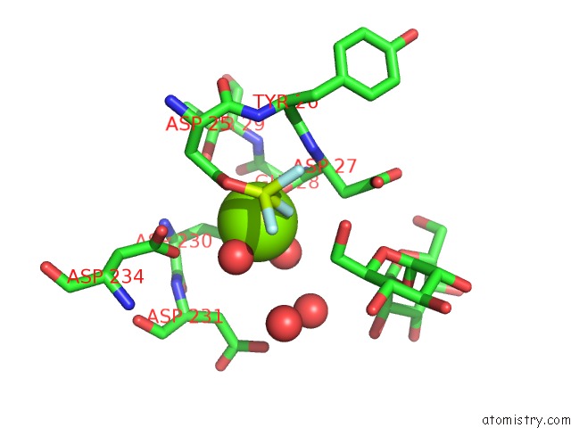
Mono view
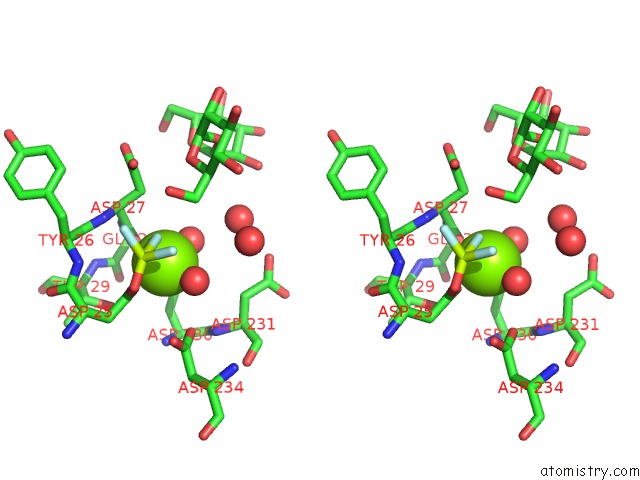
Stereo pair view
Mono view
Stereo pair view
|
|
A full contact list of Magnesium with other atoms in the Mg binding
site number 1 of Structure of C. Albicans Trehalose-6-Phosphate Phosphatase C-Terminal Domain within 5.0Å range:
|
Magnesium binding site 2 out of 2 in 5dxi
Go back to
Magnesium binding site 2 out
of 2 in the Structure of C. Albicans Trehalose-6-Phosphate Phosphatase C-Terminal Domain
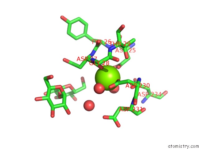
Mono view
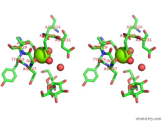
Stereo pair view
Mono view
Stereo pair view
|
|
A full contact list of Magnesium with other atoms in the Mg binding
site number 2 of Structure of C. Albicans Trehalose-6-Phosphate Phosphatase C-Terminal Domain within 5.0Å range:
|
Reference:
Y.Miao,
J.L.Tenor,
D.L.Toffaletti,
E.J.Washington,
J.Liu,
W.R.Shadrick,
M.A.Schumacher,
R.E.Lee,
J.R.Perfect,
R.G.Brennan.
Structures of Trehalose-6-Phosphate Phosphatase From Pathogenic Fungi Reveal the Mechanisms of Substrate Recognition and Catalysis. Proc.Natl.Acad.Sci.Usa V. 113 7148 2016.
ISSN: ESSN 1091-6490
PubMed: 27307435
DOI: 10.1073/PNAS.1601774113
Page generated: Sun Sep 29 03:26:30 2024
ISSN: ESSN 1091-6490
PubMed: 27307435
DOI: 10.1073/PNAS.1601774113
Last articles
Zn in 9MJ5Zn in 9HNW
Zn in 9G0L
Zn in 9FNE
Zn in 9DZN
Zn in 9E0I
Zn in 9D32
Zn in 9DAK
Zn in 8ZXC
Zn in 8ZUF