Magnesium »
PDB 5dt3-5e79 »
5e4r »
Magnesium in PDB 5e4r: Crystal Structure of Domain-Duplicated Synthetic Class II Ketol-Acid Reductoisomerase 2IA_KARI-Dd
Enzymatic activity of Crystal Structure of Domain-Duplicated Synthetic Class II Ketol-Acid Reductoisomerase 2IA_KARI-Dd
All present enzymatic activity of Crystal Structure of Domain-Duplicated Synthetic Class II Ketol-Acid Reductoisomerase 2IA_KARI-Dd:
1.1.1.86;
1.1.1.86;
Protein crystallography data
The structure of Crystal Structure of Domain-Duplicated Synthetic Class II Ketol-Acid Reductoisomerase 2IA_KARI-Dd, PDB code: 5e4r
was solved by
J.K.B.Cahn,
S.Brinkmann-Chen,
A.R.Buller,
F.H.Arnold,
with X-Ray Crystallography technique. A brief refinement statistics is given in the table below:
| Resolution Low / High (Å) | 103.88 / 1.94 |
| Space group | P 43 2 2 |
| Cell size a, b, c (Å), α, β, γ (°) | 103.882, 103.882, 142.074, 90.00, 90.00, 90.00 |
| R / Rfree (%) | 15.8 / 18 |
Magnesium Binding Sites:
The binding sites of Magnesium atom in the Crystal Structure of Domain-Duplicated Synthetic Class II Ketol-Acid Reductoisomerase 2IA_KARI-Dd
(pdb code 5e4r). This binding sites where shown within
5.0 Angstroms radius around Magnesium atom.
In total 2 binding sites of Magnesium where determined in the Crystal Structure of Domain-Duplicated Synthetic Class II Ketol-Acid Reductoisomerase 2IA_KARI-Dd, PDB code: 5e4r:
Jump to Magnesium binding site number: 1; 2;
In total 2 binding sites of Magnesium where determined in the Crystal Structure of Domain-Duplicated Synthetic Class II Ketol-Acid Reductoisomerase 2IA_KARI-Dd, PDB code: 5e4r:
Jump to Magnesium binding site number: 1; 2;
Magnesium binding site 1 out of 2 in 5e4r
Go back to
Magnesium binding site 1 out
of 2 in the Crystal Structure of Domain-Duplicated Synthetic Class II Ketol-Acid Reductoisomerase 2IA_KARI-Dd
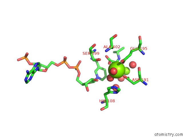
Mono view
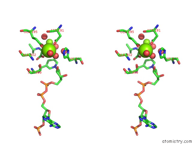
Stereo pair view
Mono view
Stereo pair view
A full contact list of Magnesium with other atoms in the Mg binding
site number 1 of Crystal Structure of Domain-Duplicated Synthetic Class II Ketol-Acid Reductoisomerase 2IA_KARI-Dd within 5.0Å range:
|
Magnesium binding site 2 out of 2 in 5e4r
Go back to
Magnesium binding site 2 out
of 2 in the Crystal Structure of Domain-Duplicated Synthetic Class II Ketol-Acid Reductoisomerase 2IA_KARI-Dd
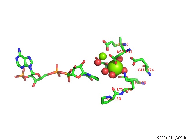
Mono view
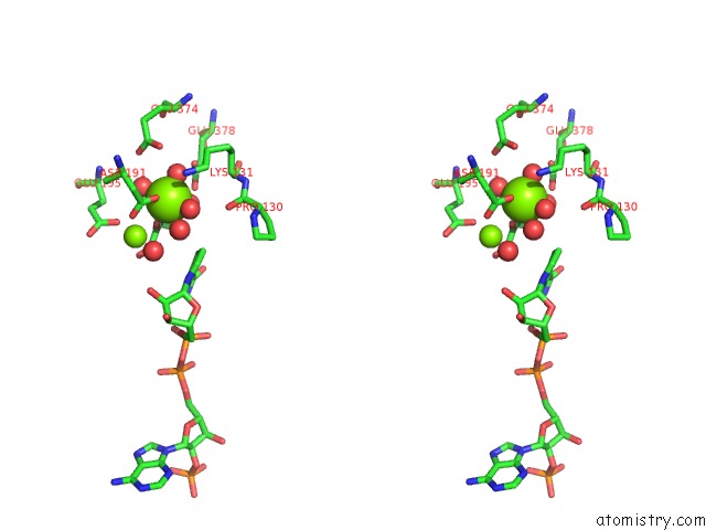
Stereo pair view
Mono view
Stereo pair view
A full contact list of Magnesium with other atoms in the Mg binding
site number 2 of Crystal Structure of Domain-Duplicated Synthetic Class II Ketol-Acid Reductoisomerase 2IA_KARI-Dd within 5.0Å range:
|
Reference:
J.K.Cahn,
S.Brinkmann-Chen,
A.R.Buller,
F.H.Arnold.
Artificial Domain Duplication Replicates Evolutionary History of Ketol-Acid Reductoisomerases. Protein Sci. V. 25 1241 2016.
ISSN: ESSN 1469-896X
PubMed: 26644020
DOI: 10.1002/PRO.2852
Page generated: Sun Sep 29 03:29:51 2024
ISSN: ESSN 1469-896X
PubMed: 26644020
DOI: 10.1002/PRO.2852
Last articles
Fe in 2YXOFe in 2YRS
Fe in 2YXC
Fe in 2YNM
Fe in 2YVJ
Fe in 2YP1
Fe in 2YU2
Fe in 2YU1
Fe in 2YQB
Fe in 2YOO