Magnesium »
PDB 5egy-5etr »
5el2 »
Magnesium in PDB 5el2: Crystal Structure of Odorant Binding Protein 1 From Anopheles Gambiae (AGAMOBP1) with Icaridin (Butan-2-Yl 2-(2-Hydroxyethyl)Piperidine-1- Carboxylate)
Protein crystallography data
The structure of Crystal Structure of Odorant Binding Protein 1 From Anopheles Gambiae (AGAMOBP1) with Icaridin (Butan-2-Yl 2-(2-Hydroxyethyl)Piperidine-1- Carboxylate), PDB code: 5el2
was solved by
C.E.Drakou,
K.E.Tsitsanou,
S.E.Zographos,
with X-Ray Crystallography technique. A brief refinement statistics is given in the table below:
| Resolution Low / High (Å) | 60.81 / 1.75 |
| Space group | P 1 21 1 |
| Cell size a, b, c (Å), α, β, γ (°) | 29.984, 68.476, 61.675, 90.00, 99.61, 90.00 |
| R / Rfree (%) | 17.8 / 22.1 |
Magnesium Binding Sites:
The binding sites of Magnesium atom in the Crystal Structure of Odorant Binding Protein 1 From Anopheles Gambiae (AGAMOBP1) with Icaridin (Butan-2-Yl 2-(2-Hydroxyethyl)Piperidine-1- Carboxylate)
(pdb code 5el2). This binding sites where shown within
5.0 Angstroms radius around Magnesium atom.
In total only one binding site of Magnesium was determined in the Crystal Structure of Odorant Binding Protein 1 From Anopheles Gambiae (AGAMOBP1) with Icaridin (Butan-2-Yl 2-(2-Hydroxyethyl)Piperidine-1- Carboxylate), PDB code: 5el2:
In total only one binding site of Magnesium was determined in the Crystal Structure of Odorant Binding Protein 1 From Anopheles Gambiae (AGAMOBP1) with Icaridin (Butan-2-Yl 2-(2-Hydroxyethyl)Piperidine-1- Carboxylate), PDB code: 5el2:
Magnesium binding site 1 out of 1 in 5el2
Go back to
Magnesium binding site 1 out
of 1 in the Crystal Structure of Odorant Binding Protein 1 From Anopheles Gambiae (AGAMOBP1) with Icaridin (Butan-2-Yl 2-(2-Hydroxyethyl)Piperidine-1- Carboxylate)
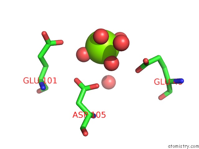
Mono view
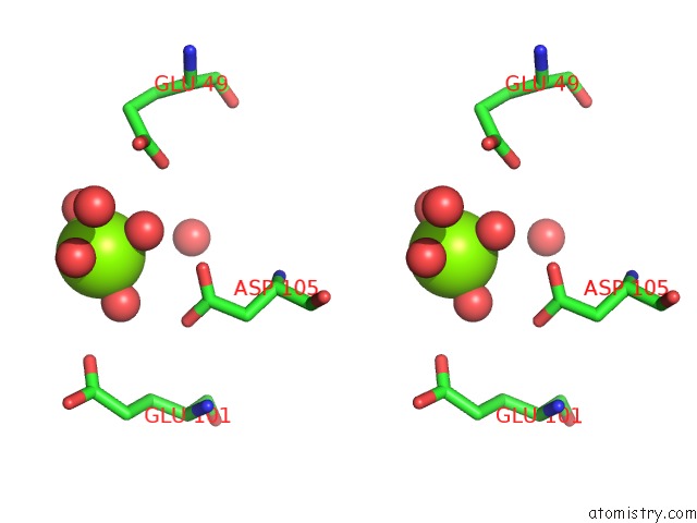
Stereo pair view
Mono view
Stereo pair view
A full contact list of Magnesium with other atoms in the Mg binding
site number 1 of Crystal Structure of Odorant Binding Protein 1 From Anopheles Gambiae (AGAMOBP1) with Icaridin (Butan-2-Yl 2-(2-Hydroxyethyl)Piperidine-1- Carboxylate) within 5.0Å range:
|
Reference:
C.E.Drakou,
K.E.Tsitsanou,
C.Potamitis,
D.Fessas,
M.Zervou,
S.E.Zographos.
The Crystal Structure of the AGAMOBP1ICARIDIN Complex Reveals Alternative Binding Modes and Stereo-Selective Repellent Recognition. Cell. Mol. Life Sci. V. 74 319 2017.
ISSN: ESSN 1420-9071
PubMed: 27535661
DOI: 10.1007/S00018-016-2335-6
Page generated: Sun Sep 29 03:50:53 2024
ISSN: ESSN 1420-9071
PubMed: 27535661
DOI: 10.1007/S00018-016-2335-6
Last articles
Fe in 6L1BFe in 6L1A
Fe in 6L32
Fe in 6L2J
Fe in 6L1X
Fe in 6KUM
Fe in 6KZT
Fe in 6KV0
Fe in 6KZS
Fe in 6KW7