Magnesium »
PDB 5fjp-5fux »
5flg »
Magnesium in PDB 5flg: Crystal Structure of the 6-Carboxyhexanoate-Coa Ligase (Biow)From Bacillus Subtilis in Complex with Amppnp
Enzymatic activity of Crystal Structure of the 6-Carboxyhexanoate-Coa Ligase (Biow)From Bacillus Subtilis in Complex with Amppnp
All present enzymatic activity of Crystal Structure of the 6-Carboxyhexanoate-Coa Ligase (Biow)From Bacillus Subtilis in Complex with Amppnp:
6.2.1.14;
6.2.1.14;
Protein crystallography data
The structure of Crystal Structure of the 6-Carboxyhexanoate-Coa Ligase (Biow)From Bacillus Subtilis in Complex with Amppnp, PDB code: 5flg
was solved by
L.Moynie,
M.Wang,
D.J.Campopiano,
J.H.Naismith,
with X-Ray Crystallography technique. A brief refinement statistics is given in the table below:
| Resolution Low / High (Å) | 41.53 / 2.04 |
| Space group | P 21 21 21 |
| Cell size a, b, c (Å), α, β, γ (°) | 49.590, 77.900, 166.000, 90.00, 90.00, 90.00 |
| R / Rfree (%) | 20.2 / 23.2 |
Magnesium Binding Sites:
The binding sites of Magnesium atom in the Crystal Structure of the 6-Carboxyhexanoate-Coa Ligase (Biow)From Bacillus Subtilis in Complex with Amppnp
(pdb code 5flg). This binding sites where shown within
5.0 Angstroms radius around Magnesium atom.
In total 4 binding sites of Magnesium where determined in the Crystal Structure of the 6-Carboxyhexanoate-Coa Ligase (Biow)From Bacillus Subtilis in Complex with Amppnp, PDB code: 5flg:
Jump to Magnesium binding site number: 1; 2; 3; 4;
In total 4 binding sites of Magnesium where determined in the Crystal Structure of the 6-Carboxyhexanoate-Coa Ligase (Biow)From Bacillus Subtilis in Complex with Amppnp, PDB code: 5flg:
Jump to Magnesium binding site number: 1; 2; 3; 4;
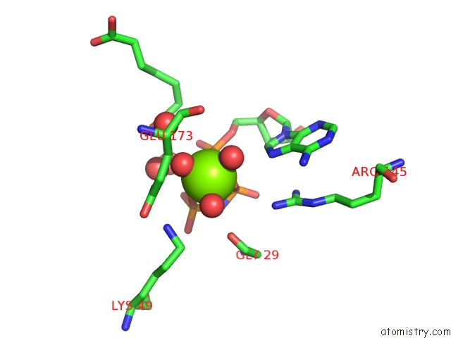
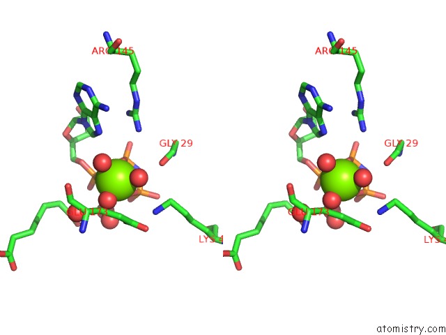
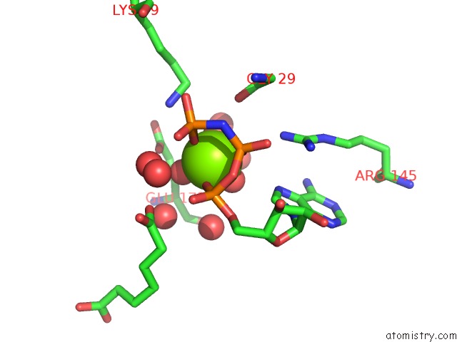
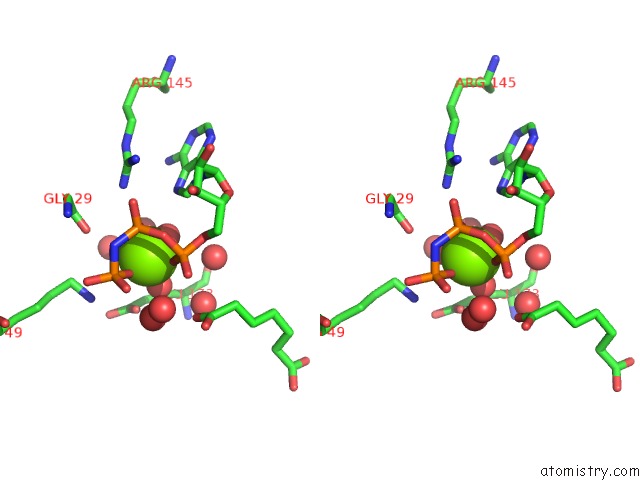
Magnesium binding site 1 out of 4 in 5flg
Go back to
Magnesium binding site 1 out
of 4 in the Crystal Structure of the 6-Carboxyhexanoate-Coa Ligase (Biow)From Bacillus Subtilis in Complex with Amppnp
Mono view
Stereo pair view
Mono view
Stereo pair view
A full contact list of Magnesium with other atoms in the Mg binding
site number 1 of Crystal Structure of the 6-Carboxyhexanoate-Coa Ligase (Biow)From Bacillus Subtilis in Complex with Amppnp within 5.0Å range:
|
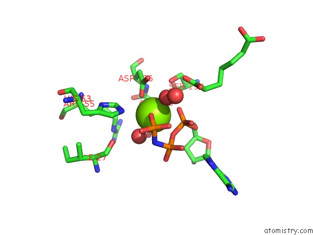
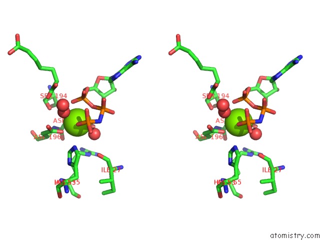
Magnesium binding site 2 out of 4 in 5flg
Go back to
Magnesium binding site 2 out
of 4 in the Crystal Structure of the 6-Carboxyhexanoate-Coa Ligase (Biow)From Bacillus Subtilis in Complex with Amppnp
Mono view
Stereo pair view
Mono view
Stereo pair view
A full contact list of Magnesium with other atoms in the Mg binding
site number 2 of Crystal Structure of the 6-Carboxyhexanoate-Coa Ligase (Biow)From Bacillus Subtilis in Complex with Amppnp within 5.0Å range:
|
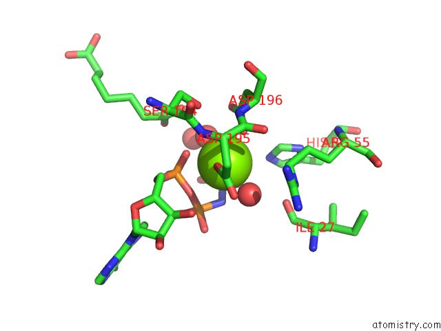
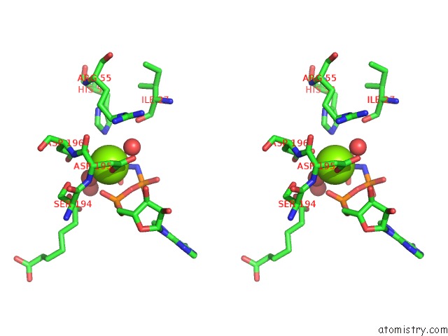
Magnesium binding site 3 out of 4 in 5flg
Go back to
Magnesium binding site 3 out
of 4 in the Crystal Structure of the 6-Carboxyhexanoate-Coa Ligase (Biow)From Bacillus Subtilis in Complex with Amppnp
Mono view
Stereo pair view
Mono view
Stereo pair view
A full contact list of Magnesium with other atoms in the Mg binding
site number 3 of Crystal Structure of the 6-Carboxyhexanoate-Coa Ligase (Biow)From Bacillus Subtilis in Complex with Amppnp within 5.0Å range:
|
Magnesium binding site 4 out of 4 in 5flg
Go back to
Magnesium binding site 4 out
of 4 in the Crystal Structure of the 6-Carboxyhexanoate-Coa Ligase (Biow)From Bacillus Subtilis in Complex with Amppnp
Mono view
Stereo pair view
Mono view
Stereo pair view
A full contact list of Magnesium with other atoms in the Mg binding
site number 4 of Crystal Structure of the 6-Carboxyhexanoate-Coa Ligase (Biow)From Bacillus Subtilis in Complex with Amppnp within 5.0Å range:
|
Reference:
M.Wang,
L.Moynie,
P.J.Harrison,
V.Kelly,
A.Piper,
J.H.Naismith,
D.J.Campopiano.
Using the Pimeloyl-Coa Synthetase Adenylation Fold to Synthesize Fatty Acid Thioesters. Nat. Chem. Biol. V. 13 660 2017.
ISSN: ESSN 1552-4469
PubMed: 28414710
DOI: 10.1038/NCHEMBIO.2361
Page generated: Sun Sep 29 04:19:42 2024
ISSN: ESSN 1552-4469
PubMed: 28414710
DOI: 10.1038/NCHEMBIO.2361
Last articles
K in 8Q46K in 8Q45
K in 8Q25
K in 8Q1Y
K in 8Q1U
K in 8Q1P
K in 8Q0Q
K in 8Q0O
K in 8Q0M
K in 8Q0J